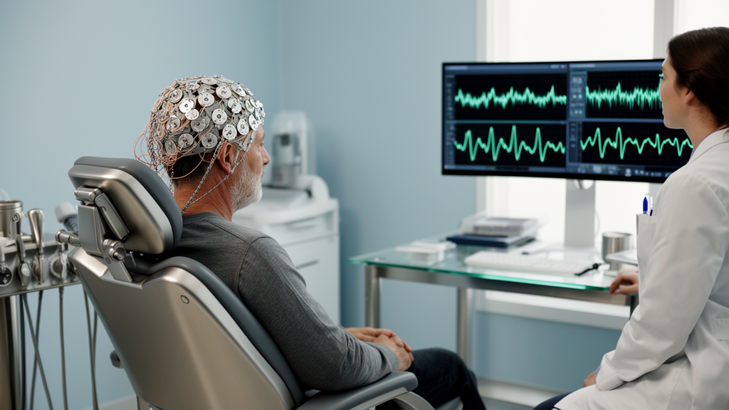
Researchers published a study in Brain Informatics in May 2026 introducing a deep learning framework designed to detect mild cognitive impairment (MCI) from electroencephalography (EEG) signals. The proposed architecture, named CNN-Res-SE-BiLSTM-BiGRU, integrates convolutional neural networks with bidirectional recurrent layers to analyze the complex temporal dynamics of brain activity. The study emphasizes not only classification accuracy but also model interpretability, aiming to address the “black box” nature of many existing deep learning models in clinical settings.
The research team utilized two distinct datasets to evaluate the model’s performance. The primary dataset consisted of resting-state EEG recordings from 27 subjects recruited in Isfahan, Iran, including 16 healthy controls and 11 individuals diagnosed with MCI. A secondary validation dataset comprised odor-evoked EEG recordings from 35 subjects, specifically using the 15 healthy controls and 7 amnestic MCI participants for binary classification. This dual-dataset approach allowed the researchers to assess the model’s generalization capabilities across different recording paradigms and electrode configurations.
The architecture combines several advanced deep learning components to capture both local and long-range temporal dependencies in EEG data. A 1D-CNN backbone with residual connections extracts localized temporal features, while Squeeze-and-Excitation (SE) modules provide channel-wise attention to emphasize informative EEG channels. Following the convolutional stages, bidirectional Long Short-Term Memory (BiLSTM) and bidirectional Gated Recurrent Unit (BiGRU) layers model long-range temporal dependencies. This hybrid design aims to overcome the limitations of traditional machine learning methods that rely on handcrafted features, which may fail to capture the non-linear nature of EEG signals.
Evaluation of the model on the primary resting-state dataset revealed significant differences in performance depending on the cross-validation protocol used. Under a subject-dependent, segment-wise five-fold cross-validation, the model achieved high accuracy of 0.974 ± 0.022 and a ROC-AUC of 0.999 ± 0.001. However, when evaluated under a strict subject-independent protocol, where data from the same subject is not shared between training and testing folds, performance dropped substantially to an accuracy of 0.560 ± 0.161 and a ROC-AUC of 0.574 ± 0.310. This contrast highlights the challenge of generalizing to unseen subjects in small resting-state cohorts.
In contrast, the model demonstrated robust and stable performance on the odor-evoked EEG dataset under subject-independent evaluation. The framework achieved an accuracy of 0.956 ± 0.051, a balanced accuracy of 0.954 ± 0.043, a ROC-AUC of 0.971 ± 0.051, and a PR-AUC of 0.934 ± 0.132. The pooled confusion matrix for this dataset showed 1,411 true negatives, 86 false positives, 28 false negatives, and 804 true positives, indicating a low false-negative rate with controlled false positives. This suggests that stimulus-locked sensory paradigms may provide more consistent signatures of early cognitive impairment than unconstrained resting-state activity.
To enhance clinical utility, the study incorporated probability calibration and specific operating threshold selection. Predicted probabilities were calibrated using temperature scaling, a post-hoc method that adjusts the model’s confidence scores. The operating threshold was selected on the validation set using Youden’s J statistic to balance sensitivity and specificity. Fixed thresholds of 0.35 and 0.50 were also reported to characterize the sensitivity-specificity trade-offs, allowing for practical tuning of the decision rule without retraining the model.
Interpretability analyses were conducted using UMAP, SHAP, and LIME to provide insight into the model’s decision-making process. UMAP visualization of the learned feature space showed moderate separation between healthy control and MCI classes, supported by a silhouette coefficient of 0.6452. SHAP analysis identified specific electrode-time-bin features as influential, with prominent contributions from prefrontal (Fp2, F8), temporal (T3, T4, T5), and occipital (O2) regions. These findings align with neurophysiological expectations, as these cortical areas are implicated in memory and executive functions often disrupted in MCI.
LIME explanations provided instance-level insights into individual predictions. For a representative healthy control segment, features such as Fp2_bin23 and F4_bin23 supported the classification, while for an MCI segment, features like Fp1_bin22 and C4_bin19 contributed to the positive prediction. These local explanations demonstrate that the model’s decisions arise from a balance of supportive and opposing contributions from specific spatiotemporal patterns, rather than relying on a single dominant feature.
Ablation studies confirmed the contribution of each architectural component to the overall performance. Removing the SE block or residual connections led to a reduction in accuracy and balanced accuracy, indicating their role in stabilizing optimization and emphasizing informative features. The most significant degradation occurred when the BiGRU component was removed, which sharply reduced the ROC-AUC and PR-AUC. This suggests that the BiGRU layer is critical for preserving class separability across unseen subjects, contributing to intrinsic discriminative power rather than merely shifting the operating point.
Baseline comparisons showed that the proposed hybrid architecture outperformed single-stream models and other CNN-recurrent variants. The strongest baseline, a CNN+BiLSTM+GRU-head model, achieved an accuracy of 0.934 ± 0.074, which was lower than the proposed model’s 0.956 ± 0.051 on the odor dataset. These results support the effectiveness of the specific combination of residual learning, SE attention, and bidirectional recurrent modeling in capturing the complex spatiotemporal dynamics of EEG signals associated with MCI.
The study acknowledges several limitations, primarily the small sample size of the primary resting-state dataset, which led to high variability in subject-independent performance. The odor-evoked dataset, while showing strong results, used only four channels and a smaller number of MCI subjects, limiting fine-grained spatial inference. The authors note that larger, multi-center cohorts are necessary to reliably quantify subject-independent generalization and stabilize calibration. Additionally, the current attention mechanism focuses on the spatial-channel dimension without explicitly modeling temporal-frequency structure, which is identified as a direction for future research.
Future work will focus on validating the model in larger cohorts and extending the attention mechanisms to capture temporal-frequency structures through cross-domain attention. The researchers aim to develop more clinically meaningful region-based explanations by mapping attributions to functional brain regions. Overall, the study demonstrates the feasibility of an EEG-based MCI screening model that combines high discrimination with calibrated decision-making and interpretability, offering a potential tool for early detection in clinical settings.
The primary resting-state EEG dataset was acquired using a 32-channel digital system at a sampling rate of 256 Hz, with nineteen active electrodes positioned according to the International 10–20 system. Each subject underwent a continuous 30-minute recording while resting with eyes closed in a quiet environment. To ensure signal quality, the researchers applied a 50 Hz notch filter to suppress power-line noise and a third-order Butterworth band-pass filter with cut-offs of 0.5–50 Hz to retain principal EEG rhythms while attenuating low-frequency drift and high-frequency noise. Segments contaminated by motion or electrode displacement were manually excluded through expert visual inspection. Following denoising, the continuous recordings were segmented into non-overlapping 6-second epochs, yielding 300 segments per subject and a total of 8,100 labeled segments across the 27-subject cohort. This preprocessing protocol was designed to preserve diagnostically relevant EEG dynamics while reducing baseline drift and muscular artifacts.
The secondary odor-evoked EEG dataset was collected to characterize neural responses to olfactory stimulation in elderly participants with different cognitive statuses. This dataset comprised recordings from 35 subjects, including 13 with Alzheimer’s disease, 7 with amnestic mild cognitive impairment, and 15 healthy controls. For the binary classification objective, the researchers excluded the Alzheimer’s disease participants and used only the healthy controls and amnestic MCI subjects. EEG signals were acquired from four electrodes at Fp1, Fz, Cz, and Pz, with Fp1 used to monitor ocular activity. Odor stimulation was delivered via a laboratory-grade olfactometer using lemon and rose in a randomized oddball design, where lemon served as the frequent stimulus with 75% probability and rose as the infrequent stimulus with 25% probability. Each trial consisted of a 2-second odor presentation followed by an 8-second rest interval. The signals were pre-processed using 0.5–40 Hz band-pass filtering, downsampling to 200 Hz, and independent component analysis for artifact removal, before being segmented into epochs from 1 second before to 2 seconds after stimulus onset.
The training strategy employed the Adam optimizer with a learning rate of 0.001 and a batch size of 16, with training conducted for a maximum of 120 epochs. To mitigate overfitting and ensure stable convergence, the researchers implemented early stopping if validation loss did not improve for 10 consecutive epochs, along with learning rate reduction on plateau. Training-fold class weights were computed exclusively from the training split to address the skewed distribution of MCI samples without using synthetic augmentation. This approach allowed the model to maintain stable optimization while accounting for the relatively fewer MCI cases in the dataset. The use of validation-only calibration and threshold selection ensured that test data were not used for tuning, reducing the risk of optimistic bias in the reported performance metrics.
The interpretability analyses were conducted on the primary dataset under the segment-wise subject-dependent setting, where the full multichannel electrode configuration supports electrode-level attribution. The SHAP analysis identified a consistent set of influential electrodes spanning frontal, prefrontal, temporal, and posterior regions. Specifically, the temporal electrodes T3, T4, and T5, which show prominent SHAP contributions, are anatomically located over the lateral temporal cortex and are functionally linked to memory-related processes. These regions lie in close proximity to the medial temporal lobe, which includes the hippocampus, one of the earliest structures affected during the progression of MCI and Alzheimer’s disease. The identification of prefrontal electrodes Fp2, F4, and F8, along with the occipital electrode O2, underscores the neurophysiological relevance of the model’s learned features, as these sites correspond to cortical regions implicated in cognitive functions frequently disrupted in MCI. The prefrontal cortex is involved in attentional control and executive function, while the occipital cortex supports visual integration, both of which show altered EEG patterns in individuals with MCI.
The study notes that the current attention mechanism is primarily applied in the spatial-channel dimension, without explicitly modeling the temporal-frequency structure. Recent advancements in EEG analysis suggest that emphasizing spectral and time-frequency characteristics can improve feature representation for neurological disorder classification. Accordingly, the authors identify the development of cross-domain attention strategies, such as joint channel–temporal–frequency attention, as a direction for future research to better capture complementary spatial, temporal, and spectral biomarkers. Additionally, the interpretability analysis, while providing electrode-level insights, is limited by its generation under a subject-dependent protocol. Future work should advance toward more clinically meaningful region and topography-based explanations by mapping attributions to functional brain regions and network-level descriptions, thereby enhancing the transparency and clinical reliability of the model’s decision-making process.
Subject of Research: A deep hybrid CNN-BiLSTM-BiGRU architecture with explainability for mild cognitive impairment detection using EEG
Article Title: A deep hybrid CNN-BiLSTM-BiGRU architecture with explainability for mild cognitive impairment detection using EEG
Article References: Tokdar, A., Agarwal, L., Chatterjee, S., & Sukriti (2026). A deep hybrid CNN-BiLSTM-BiGRU architecture with explainability for mild cognitive impairment detection using EEG. Brain Informatics, 13(1), Article 17. https://doi.org/10.1186/s40708-026-00302-4
Image Credits: AI Generated
DOI: 10.1186/s40708-026-00302-4
Keywords: deep, hybrid, CNN-BiLSTM-BiGRU, architecture, explainability, mild, cognitive, impairment, detection, scientific research
News Source: Denise Maddox. (November 11, 2026). Hybrid AI Model Detects Mild Cognitive Impairment from EEG. Scienmag.