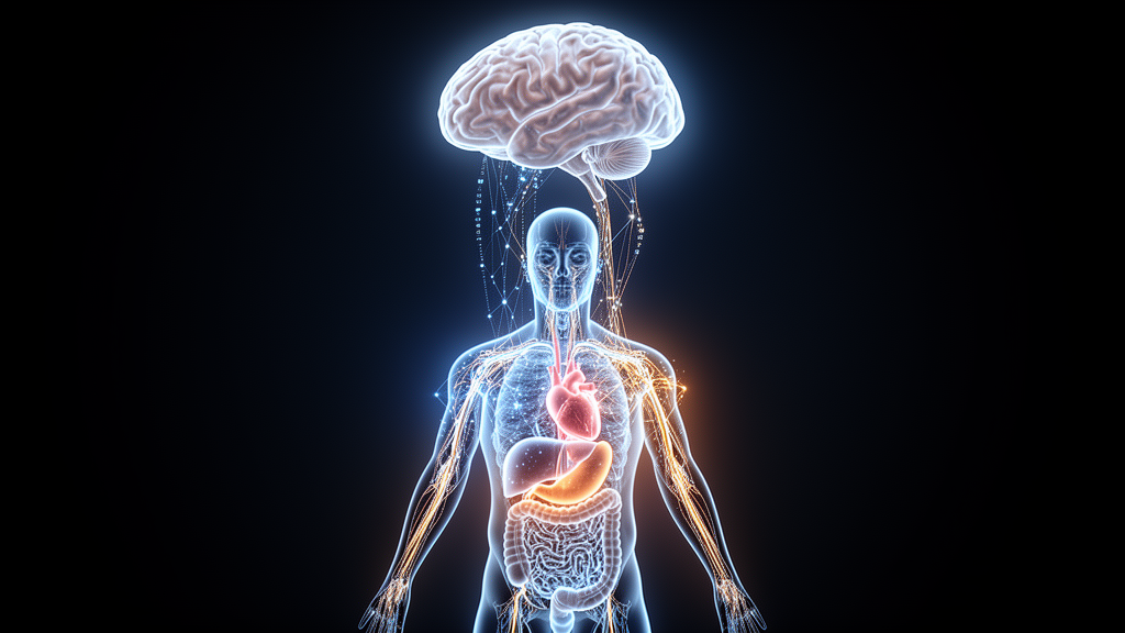
For decades, the study of how genes shape the brain has been a strangely solitary enterprise. Neuroscientists scanned brains, cardiologists scanned hearts, and skeletal biologists mapped bones, each field extracting quantitative traits from medical images and hunting for genetic variants that explain them. Now, in a perspective published in Nature Reviews Neurology, Zirui Fan and Bingxin Zhao of the University of Pennsylvania argue that this organ-by-organ approach has reached its limits. The emerging discipline they call brain-focused multi-organ imaging genetics treats the brain as one node in a whole-body network, and it is already uncovering genetic connections between neurological disorders and imaging features of the heart, retina, liver, vasculature and skeleton that no single-organ study could have detected.
The technical foundation of the field is the imaging-derived phenotype, or IDP: a quantitative measurement extracted from a medical image that captures structural or functional features of tissue. In the brain, IDPs include cortical thickness, regional volumes, white matter microstructure measured by diffusion MRI, and intrinsic functional networks derived from resting-state scans. In the body, they span cardiac chamber volumes and myocardial mass from cardiac MRI, visceral and liver fat from abdominal imaging, retinal layer thicknesses and vessel tortuosity from optical coherence tomography, and carotid intima-media thickness from ultrasound. When hundreds of thousands of individuals carry both genome-wide genetic data and whole-body multimodal imaging, researchers can run genome-wide association studies, or GWAS, on thousands of these phenotypes simultaneously, mapping the genetic determinants of every organ in a single population.
The pivotal resource enabling this shift is the UK Biobank, which combines deep phenotyping and genomic data for roughly half a million participants, including whole-body MRI, brain MRI and retinal imaging for an imaging enhancement of 100,000 people. Landmark GWAS of brain imaging phenotypes in this cohort, published in Nature in 2018, established a foundational genetic map of human brain structure and function, and subsequent expanded analyses have pushed the number of mapped traits into the tens of thousands. Parallel efforts have extracted genetic architecture from cardiac MRI in 36,000 individuals, abdominal MRI organ traits via deep learning, liver fat through machine learning, and the human skeletal form. Complementary cohorts such as CHIMGEN in China, IMAGEN in adolescent psychiatry, the ABCD study of child brain development and the ENIGMA consortium spanning more than 40 countries provide cross-ethnic and cross-geographic depth, while whole-genome sequencing and exome data from hundreds of thousands of UK Biobank participants extend discovery from common variants to rare copy number variants and coding mutations.
What has emerged from these datasets is a distributed whole-body genetic architecture underlying neurological and neuropsychiatric conditions. Studies of heart-brain connections using cardiac and brain MRI found systematic phenotypic and genetic correlations between cardiac structure and brain imaging traits, laying the groundwork for the brain-body perspective. Multi-organ imaging in UK Biobank participants demonstrated a heart-brain-liver axis, with associations spanning all three organs. Eye-brain imaging genetics revealed shared genetic architecture between retinal and brain traits, showing that accessible peripheral imaging can inform brain biology. For Alzheimer disease, multi-trait analyses have identified shared genetic loci between the disorder and cardiovascular traits, and genetic signatures of heart function overlap with dementia risk. For schizophrenia, shared genetic architecture links the illness to common peripheral organ imaging phenotypes, and retinal thinning has been connected to neuroinflammatory pathways associated with genetic susceptibility to the disorder.
Statistical association alone, however, does not establish mechanism, and the field has developed a principled three-step analytical pipeline to move from correlation towards causation. The first step is causal gene nomination: statistical fine-mapping narrows GWAS loci to candidate causal variants, colocalization tests assess whether associations between two traits at the same genomic locus are likely driven by a shared causal variant, and integration with quantitative trait loci resources such as the GTEx atlas of genetic regulatory effects across dozens of human tissues, brain eQTL and pQTL fine-mapping, and single-cell transcriptomic atlases of the human brain nominates the genes and cell types through which variants act. The second step builds organ-enriched network modules: network propagation expands seed genes into broader functional modules through protein-protein interaction networks, and genomic structural equation modelling characterizes the multivariate genetic architecture linking organ systems. The third step is cross-field validation, testing whether nominated genes and pathways hold up across independent datasets, model systems and experimental platforms.
Mendelian randomization, which uses genetic variants as instrumental variables to investigate potential causal relationships between an exposure and an outcome, has become the workhorse for interrogating cross-organ connections. Bidirectional analyses have supported causal links between brain imaging-derived phenotypes and stroke, including thalamic nuclei volumes and stroke risk, between aortic distensibility and brain white matter hyperintensities, between kidney damage and cortical structure, and between structural brain changes and risks of Alzheimer disease, Parkinson disease, amyotrophic lateral sclerosis, epilepsy, migraine and major psychiatric disorders. Drug target Mendelian randomization has begun to reshape clinical translation: lower activity of the cholesteryl ester transfer protein, or CETP, has been associated with reduced dementia risk, and evidence from drug target Mendelian randomization is being used to repurpose existing drugs for prevention of vascular dementia. The approach also clarifies long-standing observational puzzles, such as the bidirectional relationship between body mass index and Alzheimer disease, where genetic analyses suggest that lower BMI by midlife may reflect earliest manifestations of the disease rather than a protective effect.
Biological ageing provides a unifying quantitative framework for the whole-body view. Studies modelling heterogeneous ageing across ten brain and body systems in UK Biobank have characterized ageing patterns across chronic diseases and demonstrated their predictive value for mortality risk, while the genetic architecture of biological age has been mapped in nine human organ systems. Imaging-based organ-specific ageing clocks predict human diseases and mortality, organ-specific proteomic ageing clocks track health and longevity across diverse populations, and brain charts for the human lifespan provide normative references against which deviations can be quantified. These clocks capture diversity and disparities in ageing and dementia across geographically diverse populations, and they connect brain ageing with immune system ageing through plasma proteomics, reinforcing the idea that brain health is inseparable from systemic biology.
The scale and complexity of multi-organ imaging genetics now exceed what any individual laboratory can manage, and the authors argue that agentic artificial intelligence co-scientists will be essential for realizing an integrated understanding of brain health and disease. Recent demonstrations show what such systems can do: an AI co-scientist that generates, debates and prioritizes scientifically testable hypotheses in collaboration with human researchers, a virtual lab of AI agents that designed experimentally validated SARS-CoV-2 nanobodies, multi-agent frameworks that autonomously select and coordinate analytical tools for biomedical data-analysis workflows, and general-purpose biomedical AI agents that integrate heterogeneous data resources and execute complex multistep scientific workflows. In imaging genetics, such systems could orchestrate the entire discovery pipeline, from GPU-accelerated GWAS on thousands of quantitative phenotypes, through fine-mapping and colocalization across organ systems, to network module construction and cross-field validation, while serving as collaborative infrastructure for the whole field rather than tools for individual researchers alone.
The authors are careful to note that AI cannot do good science without humans, and that reproducible brain-wide association studies require thousands of individuals, a constraint that motivates large-scale collaborative imaging research. Challenges remain in phenotypic and genetic heterogeneity, in the specificity of signals from minimal phenotyping, in diversity of genomic research, and in the statistical enemies of reliable clinical prediction models. Yet the trajectory is clear. Clinical practice guidelines for brain-heart multimorbidity management, scientific statements on brain health across the lifespan from the American Heart Association, and reviews of bidirectional brain-heart interactions in health and disease signal that clinical care is beginning to move beyond organ boundaries. As multi-organ imaging genetics matures, the genetic determinants of brain disorders are being understood not in isolation but as part of a whole-body architecture, and the integrated view of brain, body and brain diseases that the field promises is no longer a distant aspiration but an active research program with a concrete roadmap.
Subject of Research: Multi-organ imaging genetics reveals a whole-body genetic architecture of brain diseases
Article Title: Multi-organ imaging genetics: towards an integrated view of brain, body and brain diseases
Article References: Fan, Z., & Zhao, B. (2026). Multi-organ imaging genetics: towards an integrated view of brain, body and brain diseases. Nature Reviews Neurology. https://doi.org/10.1038/s41582-026-01276-x
Image Credits: AI Generated
DOI: 10.1038/s41582-026-01276-x
Keywords: Multi-organ, imaging, genetics, towards, integrated, view, brain, body, diseases, scientific research
News Source: Juliet Wilcox. (October 19, 2026). How Whole-Body Imaging Genetics Is Rewriting the Map of Brain Disease. Scienmag.