Studying the developing embryo of the ‘frilled dragon’ lizard, UNIGE researchers reveal that physical forces, rather than a genetic program, generate the characteristic folds of its spectacular collar.
Credit: © UNIGE, A.Debry/S.Montandon/M.Milinkovitch
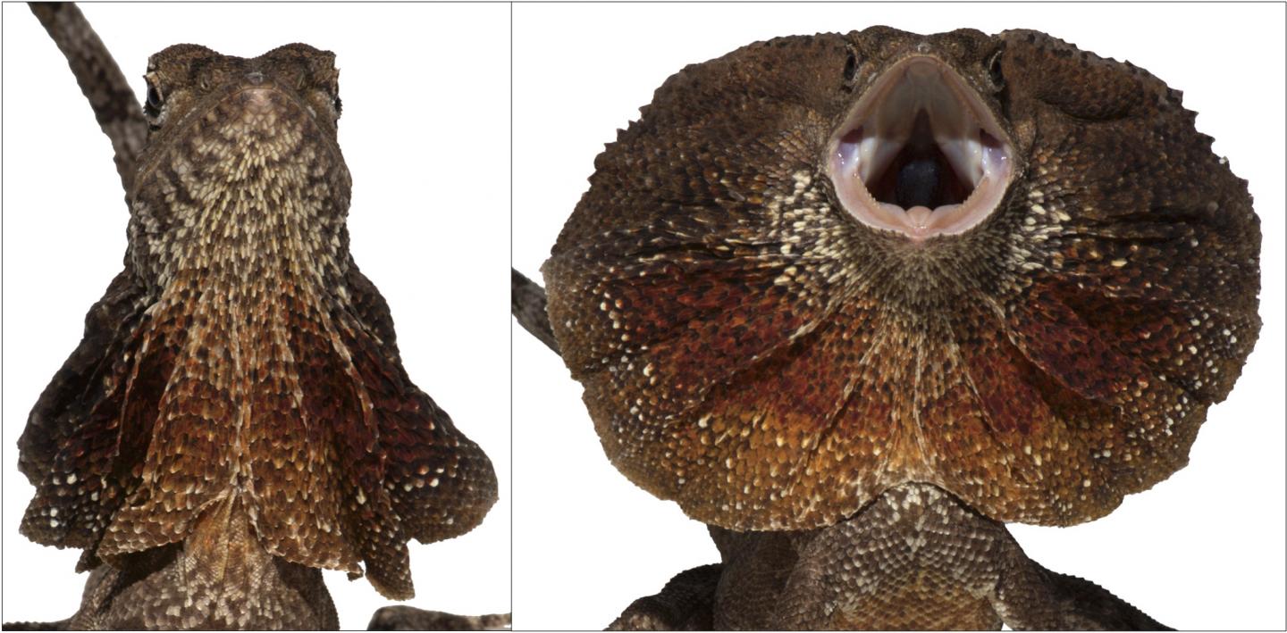
The frilled dragon exhibits a distinctive large erectile ruff. This lizard usually keeps the frill folded back against its body but can spread it as a spectacular display to scare off predators. Researchers at the University of Geneva (UNIGE), Switzerland, and the SIB Swiss Institute of Bioinformatics report in the journal eLife that an ancestral embryonic gill of the dragon embryo turns into a neck pocket that expands and folds, forming the frill. The researchers then demonstrate that this robust folding pattern emerges from mechanical forces during the homogeneous growth of the frill skin, due to the tensions resulting from its attachment to the neck and head.
In Jurassic Park, while the computer programmer Dennis Nedry attempts to smuggle dinosaur embryos off the island, he gets attacked and killed by a mid-sized dinosaur that erects a frightening neck frill. This fictional dinosaur is clearly inspired from a real animal known as the ‘frilled dragon’, that lives today in northern Australia and southern New Guinea. These lizards, also known as Chlamydosaurus kingii, have a large disc of skin that sits around their head and neck. This frill is usually folded back against the body, but can spread in a spectacular fashion to scare off predators and competitors. Folding of the left and right sides of the frill occurs at three pre-formed ridges. But, it remains unclear which ancestral structure evolved to become the dragon’s frill, and how the ridges in the frill form during development.
Recycling gills
A multidisciplinary team led by Michel Milinkovitch, Professor at the Department of Genetics and Evolution of the UNIGE Faculty of Science and Group Leader at the SIB Swiss Institute of Bioinformatics, shows today that the dragon’s frill, as well as bones and cartilages that support it, develop from the branchial arches. These are a series of bands of tissue in the embryo that evolved to become the gill supports in fish, and that now give rise to multiple structures in the ear and neck of land vertebrates. In most species, the second branchial arch will eventually fuse with the arches behind it. But in the frilled dragon, this arch continues instead to expand, leading to the formation of the dragon’s spectacular frill. “These changes in the development of gill arches highlight how evolution is able to “recycle” old structures into new forms playing different roles,” enthuses Michel Milinkovitch.
Mechanical process rather than molecular genetic signal
As the frill develops, the front side of the skin forms three successive folds, which make up the pre-formed ridges. Studying the formation of these ridges, the Swiss team reveals that they do not emerge from increased growth at the folding sites, but from physical forces – whereby the growth of the frill is constrained by its attachment to the neck. This causes the top layer to buckle, creating the folds of the frill. “We then simulated this process in a computer model and discovered that we could trace how the folds develop in the frills of the real lizard embryos,” continues Michel Milinkovitch.
###
These results provide further evidence that physical processes, as well as genetic programs, can shape tissues and organs during an embryo’s development.
Media Contact
Michel Milinkovitch
[email protected]
Related Journal Article
http://dx.


