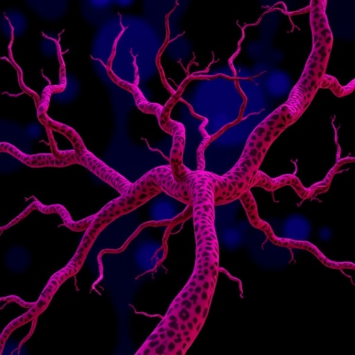
Pericytes, the elongated mural cells that wrap themselves around the smallest blood vessels in the body, have long been treated as supporting actors in cancer biology. A comprehensive review published in Advanced Science now argues that they deserve top billing. Written by Ziheng Guo of West China Hospital, Sichuan University, and Yihai Cao of Karolinska Institutet, the analysis assembles a decade of experimental evidence showing that these perivascular cells orchestrate nearly every stage of the metastatic cascade — from the moment a cancer cell squeezes into the bloodstream to its preparation of distant tissues for colonization. The central message is paradoxical and striking: the very cells that stabilize blood vessels and hold tumors in check when anchored in place can become some of cancer’s most effective accomplices once they detach, change identity, and begin secreting the molecular signals that help tumors spread.
Structurally, pericytes sit within the basement membrane that surrounds the endothelial cells lining capillaries and post-capillary venules. In healthy tissue, this intimate anatomical position allows them to perform a range of physiological duties: they regulate capillary diameter and blood flow, prevent vascular leakiness, maintain the blood-brain barrier, and modulate inflammatory cell traffic across the vessel wall. They are also remarkably plastic, retaining mesenchymal stem cell-like properties that permit differentiation into fibroblasts, myocytes, adipocytes, and osteocytes. Marker-based identification remains a persistent challenge because no single molecule defines all pericytes; researchers typically rely on combinations of platelet-derived growth factor receptor beta (PDGFR-β), the chondroitin sulfate proteoglycan NG2, alpha-smooth muscle actin, desmin, CD13, and, more recently, markers such as Gli1 and Tbx18. This heterogeneity — shaped by tissue of origin, which ranges from mesothelium to neural crest to hematopoietic myeloid progenitors — complicates every attempt to generalize about their behavior in disease.
Within tumors, the relationship between pericytes and endothelial cells frequently breaks down. Rapidly growing tumors become hypoxic, driving hypoxia-inducible factor 1 alpha-mediated expression of vascular endothelial growth factor, which promotes disorganized, leaky angiogenesis and can even strip pericytes from microvessels. Genetic mutations amplify the problem. In epithelial cancers carrying KRAS mutations — pancreatic ductal adenocarcinoma, hepatocellular carcinoma, colorectal cancer, and non-small cell lung carcinoma — activation of the transcription factor Forkhead Box C2 upregulates angiopoietin 2, which binds the Tie2 receptor on endothelial cells and actively repels pericytes from the vessel wall. Because the Ang1/Ang2 pair acts on the same receptor with opposite effects — Ang1 stabilizes pericyte coverage while Ang2 ablates it — the balance of these ligands becomes a molecular switch determining how leaky and invasion-prone a tumor’s vasculature will be.
Perhaps the most counterintuitive finding summarized in the review concerns anti-pericyte therapy. Pharmacological ablation of pericytes using drugs that block PDGFR-β signaling, such as imatinib and sunitinib, has been shown in preclinical models to promote rather than suppress metastasis. In a genetic mouse model engineered to selectively deplete NG2-positive pericytes, pericyte loss triggered tumor hypoxia, epithelial-to-mesenchymal transition, and activation of the c-Met pathway, fueling breast cancer cell intravasation into blood vessels. Clinical data mirror these observations: in several cancer types, low pericyte coverage of tumor vessels correlates inversely with patient survival. The interpretation is that intact pericyte coating forms part of a three-layered vascular barrier — endothelium, perivascular cell layer, and basement membrane — that metastasizing cells must breach. When pericytes are stripped away, that barrier collapses, opening gates for tumor cells entering and exiting the circulation.
The review details the sequential choreography of intravasation with unusual precision. A metastatic cancer cell must first degrade the local basement membrane, then survive the loss of pericyte coverage, and finally pry open the tight junctions between endothelial cells to slip into the bloodstream. Pericytes facilitate the first step through signaling: when stimulated by PDGF-BB, they dramatically upregulate interleukin-33 — the most strongly induced gene in genome-wide profiling experiments — and increase production of matrix metalloproteinase-9 by more than 200-fold. MMP-9 digests the basement membrane surrounding microvessels, while IL-33 recruits and converts macrophages toward an M2-like, tumor-promoting phenotype. These IL-33-activated macrophages in turn release CXCL3, which drives cancer-associated fibroblasts into contractile myofibroblasts through the CXCR2 receptor, a cascade shown in pancreatic cancer models to accelerate metastatic spread through what the authors describe as a hijacked cancer-escape mechanism.
Single-cell RNA sequencing has now revealed pericyte subpopulations with remarkably specific pro-metastatic functions. In both human and mouse colorectal cancers, a subset of pericytes expressing nicotinamide N-methyltransferase, or NNMT, is strongly associated with metastatic potential. These NNMT-positive pericytes secrete the chemokine CXCL5, which promotes the formation of circulating tumor cell–neutrophil clusters — multicellular aggregates that survive better in the bloodstream and seed metastases more efficiently than single cancer cells. When researchers deleted NNMT specifically in pericytes, cluster formation and metastasis were suppressed; blocking the CXCL5/CXCR2 signaling axis produced the same effect. In patients, high numbers of NNMT-positive pericyte-coated vessels corresponded to detectable CTC-neutrophil clusters and served as a poor prognostic marker. Another subpopulation, defined by high expression of the transcription factor TCF21, promotes colorectal cancer metastasis by remodeling the perivascular extracellular matrix, while endosialin-expressing pericytes facilitate metastatic dissemination through contact-dependent mechanisms without affecting primary tumor growth.
Pericyte plasticity adds yet another layer to the story. Genetic lineage tracing of NG2-positive pericytes has demonstrated that, once detached from tumor microvessels, these cells can differentiate into cancer-associated fibroblasts and myofibroblasts — a process termed pericyte-to-fibroblast transition, or PFT. The resulting stromal cells reinforce the dense, fibrotic stroma characteristic of tumors such as pancreatic ductal adenocarcinoma, where fibrotic tissue can constitute 80 to 90 percent of the tumor mass and forms what researchers call a “fibrotic fortress” that shields cancer cells from drugs. In PDAC, most vascular pericyte-endothelial interactions are lost due to the near-universal KRAS mutations, while an aberrant alpha-SMA-positive pericyte population emerges that behaves more like a myofibroblast than a vascular support cell. A rare subpopulation called pericyte stem cells, barely detectable in healthy tissue but abundant in PDAC, further suppresses antitumor immunity by differentiating into Ly6G-positive myeloid-derived suppressor cells, thereby driving resistance to anti-PD1 immunotherapy while directly stimulating cancer cell proliferation.
The immune dimension of pericyte behavior extends well beyond the pancreas. Glioblastoma pericytes acquire immunosuppressive phenotypes by producing interleukins 1, 6, 10, 12, and 26, while simultaneously reducing IL-2 availability and secreting TGF-beta and hepatocyte growth factor to inactivate cytotoxic CD8-positive T cells. Pericytes can also upregulate the checkpoint molecules PD-L1 and PD-L2, inducing a state of T cell anergy, and in coculture experiments they profoundly inhibit T cell activation, proliferation, and production of interferon-gamma and tumor necrosis factor alpha. Significantly, this immunosuppressive state appears reversible: in mouse melanoma models, forcing pericytes to switch from an immature “synthetic” phenotype back to a differentiated state alleviated immune suppression and sensitized tumors to adoptive T cell therapy, leading to tumor regression. This finding suggests that pericyte phenotype manipulation could become a strategy for converting immunotherapy-resistant tumors into responsive ones.
Perhaps most alarming is the role pericytes play before cancer cells ever arrive at distant organs. Primary tumors release signaling molecules and exosomes into the circulation that precondition remote tissues, creating pre-metastatic niches receptive to future colonization. Lineage-tracing studies in metastatic melanoma and rhabdomyosarcoma models show that pericytes in the lung migrate away from vessels, downregulate their canonical markers, gain proliferative and migratory capacity, and begin depositing extracellular matrix proteins such as fibronectin — essentially laying the scaffold on which arriving metastatic cells will grow. Tumor-derived factors upregulate the transcription factor Kruppel-like factor 4 in these perivascular cells, and KLF4-dependent perivascular cell plasticity has been shown to be necessary for pre-metastatic niche formation and metastasis. Even lung cancer stem cells appear to exploit this system, generating pericyte-like cells that enhance trans-endothelial migration and facilitate brain metastasis.
The therapeutic implications of this body of work are double-edged. Pericytes lack a single specific molecular target, display enormous heterogeneity, and exist in every vascularized tissue, so indiscriminate targeting risks damaging healthy vasculature — and, given their barrier function, may even accelerate metastasis. Yet the identification of pro-metastatic subpopulations such as NNMT-positive, TCF21-high, desmin-positive, and CD13-positive pericytes offers a roadmap for selective intervention. Combination strategies look especially promising: pairing anti-VEGF agents with PDGF-targeting drugs has effectively treated tumors resistant to either approach alone, and neutralizing pericyte-mediated immune suppression could amplify the efficacy of existing immunotherapies. Timing remains an open question — zebrafish models show that intravasation can begin when primary tumors are still tiny, meaning metastasis may be underway before diagnosis — but the mechanistic clarity achieved in this review transforms pericytes from an obscure vascular curiosity into one of the most compelling and underexploited targets in metastatic disease research.
Subject of Research: Mechanisms by which pericytes and other perivascular cells regulate tumor angiogenesis, vascular permeability, immune suppression, intravasation, extravasation, pericyte-to-fibroblast transition, and pre-metastatic niche formation in cancer metastasis
Subject of Research: Technology and Engineering
Article Title: Mechanisms of Pericyte-Mediated Cancer Metastasis
Article References: Guo, Z., & Cao, Y. (2026). Mechanisms of Pericyte‐Mediated Cancer Metastasis. Advanced Science, 13(33), Article e00031. https://doi.org/10.1002/advs.202600031
Image Credits: AI Generated
DOI: 10.1002/advs.202600031
Keywords: pericytes, cancer metastasis, tumor microenvironment, angiogenesis, pericyte-endothelial cell interaction, pre-metastatic niche, pericyte-to-fibroblast transition, circulating tumor cells, immunosuppression, PDGF-BB, angiopoietin-2, matrix metalloproteinases
Cite Scienmag News
APA MLA Chicago
Nathaniel Bowman. (September 6, 2026). How Pericytes Drive the Deadly Spread of Cancer. Scienmag. https://scienmag.com/how-pericytes-drive-the-deadly-spread-of-cancer/
Nathaniel Bowman. “How Pericytes Drive the Deadly Spread of Cancer.” Scienmag, 6 September 2026, https://scienmag.com/how-pericytes-drive-the-deadly-spread-of-cancer/. Accessed 6 September 2026.
Nathaniel Bowman. “How Pericytes Drive the Deadly Spread of Cancer.” Scienmag. September 6, 2026. https://scienmag.com/how-pericytes-drive-the-deadly-spread-of-cancer/
Copy citation Download RIS
Tags: blood vessel regulation in tumorsblood vessel stabilization in cancerblood-brain barrier and cancercancer cell intravasationcancer cell intravasation and extravasationcancer metastasismetastatic cascade mechanismsmolecular signals in cancer spreadpericyte detachment and transformationpericyte role in tumor progressionpericyte-endothelial cell interactionspericytes as therapeutic targetsperivascular cells in cancertherapeutic targeting of pericytestumor microenvironmenttumor vascular biologytumor vascular stabilizationvascular leakiness in metastasis