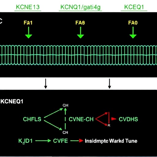
In an electrifying advancement in the field of ion channel physiology, researchers have unveiled novel insights into the intricate modulation of KCNQ1 potassium channels by their auxiliary KCNE subunits. These channels, fundamental to critical physiological processes such as cardiac rhythm maintenance and intestinal chloride secretion, orchestrate electrical signals in diverse tissues. The study sheds unprecedented light on how two members of the KCNE family—KCNE1 and KCNE3—finely tune KCNQ1’s gating mechanisms, balancing voltage-dependent activation and phosphoinositide-dependent regulation to suit the specific demands of excitable and non-excitable cells.
At the heart of this discovery lies the revelation that KCNQ1–KCNE1 and KCNQ1–KCNE3 channel complexes host not just one but two distinct PIP2 (phosphatidylinositol 4,5-bisphosphate) binding sites. PIP2, a phospholipid critical for membrane signaling, modulates KCNQ1 gating, essentially acting as an essential cofactor for channel opening. While previous research extensively characterized voltage-dependent channel activation and the role of KCNE subunits in this process, the newfound dual-site PIP2 interaction introduces a sophisticated layer of regulation previously unappreciated in this system.
The electron cryo-microscopy structural reconstructions show that KCNE1 and KCNE3 contribute directly to a previously overlooked PIP2 binding site located at the interface critical for coupling the voltage-sensor domain (VSD) to the pore domain. This novel site encompasses residues that have historically eluded functional annotation but now emerge as pivotal players driving the channel’s response to both membrane voltage and cellular phosphoinositide dynamics.
.adsslot_NL5mO8A3yv{ width:728px !important; height:90px !important; }
@media (max-width:1199px) { .adsslot_NL5mO8A3yv{ width:468px !important; height:60px !important; } }
@media (max-width:767px) { .adsslot_NL5mO8A3yv{ width:320px !important; height:50px !important; } }
ADVERTISEMENT
This dual modulation is particularly fascinating given the distinct physiological roles of KCNE1 and KCNE3. With KCNE1, the interaction strengthens KCNQ1’s affinity for PIP2, enhancing the channel’s resistance to downregulation by G protein-coupled receptor (GPCR) signaling. This stabilizing effect reinforces the traditionally voltage-dependent gating essential for generating the slow-delayed rectifier potassium current (IKs) in excitable cardiac myocytes, where precise timing is critical for cardiac repolarization and rhythm stability.
In contrast, KCNE3 turns the paradigm on its head by converting KCNQ1 into a voltage-insensitive, PIP2-gated channel. This conversion effectively places KCNQ1 function under the direct control of GPCR-driven PIP2 metabolism. In certain epithelial and non-excitable cells, such as those lining the intestine, this arrangement supports ion homeostasis by permitting channel activation independent of voltage fluctuations, favoring a biochemical modulation mode adapted to those tissues’ environmental demands.
From a physiological viewpoint, this fine-tuning mechanism crafts a versatile toolkit for cells to tailor channel behavior to their unique electrochemical environments. The same core channel protein, KCNQ1, through selective pairing with KCNE subunits, unlocks a spectrum of gating modes ranging from strict voltage dependence to biochemical gating governed by lipid signaling. This adaptability underscores a remarkable evolutionary strategy enabling a single ion channel gene to fulfill multiple roles in disparate tissues.
Importantly, the study integrates structural biology with functional electrophysiology and cellular signaling, constructing a holistic picture of how KCNE1/3 subunits influence KCNQ1 gating. The high-resolution cryo-EM data allowed precise mapping of PIP2 binding sites, illuminating how subunit-specific interactions reconfigure the channel to modulate its sensitivity to PIP2 and consequently, its gating behavior. It confronts previous ambiguities regarding the role of PIP2 and accessory subunits in KCNQ1 regulation.
The implications for cardiac physiology are profound. KCNE1’s ability to bolster PIP2 affinity and confer robustness against GPCR-mediated inhibition equips cardiac cells with a stable potassium current essential for normal heart rhythm. Disruptions in this balance are linked to arrhythmias and long QT syndrome, conditions that threaten life. By clarifying molecular determinants of KCNE1’s modulatory effects, this study opens avenues to targeted drug design aimed at selectively modulating IKs without affecting other KCNQ1 functions.
Likewise, the elucidation of KCNE3’s gating conversion in epithelial cells illuminates pathophysiological mechanisms underlying disorders like cystic fibrosis, where chloride secretory defects contribute to disease. The coupling of KCNQ1 gating to GPCR and PIP2 signaling dynamics suggests new potential targets for modulating ion transport therapeutically in epithelial tissues.
Beyond physiology and disease, this work exemplifies how ion channel auxiliary subunits serve as molecular rheostats, precisely adjusting the functional repertoire of ion channels. It invites a reevaluation of channelopathies through the lens of subunit diversity and lipid regulation, expanding the conceptual framework of membrane excitability and signaling.
Technically, achieving these insights hinged on resolving ambiguities in previously reported KCNQ1–KCNE3 structures and unveiling subtle, functionally critical lipid interaction sites. The identification of the secondary PIP2 site required integrating biochemical, structural, and mutagenesis data to pinpoint residues contributing to channel gating coupling. Such integrative approaches set new standards for channel biophysics investigations.
Moreover, the findings illustrate the power of GPCR-coupled lipid signaling to act as a functional switch on channels long regarded as predominantly voltage-dependent. This dual gating mechanism invites further exploration of how other members of the KCNE family may similarly program KCNQ1 or related channels, hinting at an array of cell-type-specific modulatory paradigms yet to be discovered.
In sum, this research redefines our molecular understanding of KCNQ1 potassium channel regulation, revealing a nuanced interplay between voltage sensing and lipid-dependent gating orchestrated by KCNE subunits. It highlights new principles of cellular specialization where subtle molecular interactions allow a single ion channel to fulfill divergent physiological roles, adapting dynamically to its cellular milieu. These insights pave the way for precision targeting of multifunctional ion channels in a tissue-specific manner, potentially revolutionizing therapeutic approaches for cardiac arrhythmias and epithelial transport disorders.
As the field moves forward, the detailed mechanistic framework provided by this work will catalyze further studies dissecting the dynamic interactions of ion channels with membrane lipids and auxiliary proteins. The prospect of exploiting dual gating modes to design novel modulators with enhanced tissue selectivity holds promise for overcoming limitations of current ion channel pharmacology, heralding a new era in treating channelopathies with refined precision.
Subject of Research: KCNQ1 potassium channel gating modulation by KCNE1 and KCNE3 subunits and their interaction with PIP2 in GPCR-mediated cellular signaling.
Article Title: Mechanisms of KCNQ1 gating modulation by KCNE1/3 for cell-specific function
Article References:
Cui, C., Zhao, L., Kermani, A.A. et al. Mechanisms of KCNQ1 gating modulation by KCNE1/3 for cell-specific function. Cell Res (2025). https://doi.org/10.1038/s41422-025-01152-1
Image Credits: AI Generated
Tags: auxiliary subunits in ion channelscardiac rhythm regulationcryo-microscopy structural analysisdual-site PIP2 interactionexcitable and non-excitable cellsgating mechanisms in ion channelsion channel physiologyKCNE1 KCNE3 modulationKCNQ1 potassium channelsmembrane signaling phospholipidsphosphoinositide binding sitesvoltage-dependent activation mechanisms
