Credit: CDT
As it is known from the international experience with COVID-19 diagnostics, the PCR test has quite limited accuracy and a long processing time. Radiology diagnostics became the frontline area in the fight with the pandemic: sensitivity of computed tomography is around 98%.
The Moscow Center for Diagnostics and Telemedicine (CDT), under the leadership of Prof. Morozov, Chief Regional Radiology Officer of Moscow, successfully developed common operation principles of radiology departments that applied to all medical facilities, including public, private, and federal, and would allow to diagnose COVID-19 efficiently and promptly.
The infrastructure of Moscow radiology diagnostic services was prepared in advance in 2012-2018. IT provision was arranged, and city clinics were provided with CT scanners. Medical facilities connected about 1000 devices to the Unified Radiological Information Service (URIS). This service conducts a cycle of diagnostic study in a single digital space. Radiologists’ work was organized remotely.
The CDT prepared clinical recommendations for the organization process of scanning, separation of patient flows, and division of functional space of radiology departments into “green” and “red” zones. All these measures were taken to prevent the spread of the infectious disease. The CDT launched operation of the Moscow Reference Center, and introduced artificial intelligence technologies into radiologists’ workflow, etc. More details are below.
Outpatient CT centers
The Moscow government, together with the CDT, decided to urgently launch a network of outpatient CT centers (OCTC) based on city clinics in order to link patients who were treated at home with hospitals, and to provide them with available diagnostics. Also, the goals of this innovation were to reduce workload on medical facilities under the conditions of rapidly spreading disease, and to reduce mortality rates from COVID-19.
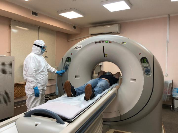
OCTCs operate on the base of outpatient facilities around the clock and perform comprehensive examination of patients providing CT scans, pulse-oximetry, ECG, examination by a physician, drawing blood for analysis, and taking swabs for PCR. Professor Morozov reported the following statistics: “In Moscow, 48 outpatient CT centers operate around the clock. From April 13th till June 20th, more than 170,000 chest CT scans were performed, more than 82,000 cases of pneumonia with COVID-19 features were detected (50% of the total number of chest CT scans). All cases of COVID-19 pneumonia have the following distribution by the degree of detected findings: CT-1 (mild) – more than 49,000 cases (60% of detected COVID-19 pneumonia); CT-2 (moderate) -more than 24,000 (29% of detected COVID-19 pneumonia); CT-3 (severe) – more than 8,000 (10% of detected COVID-19 pneumonia); CT-4 (critical) – more than 750 (1% of detected COVID-19 pneumonia)”. In addition to the above, the CDT assisted in the rapid launch of CT rooms in restructured hospitals to combat COVID-19.
Ensuring safety of healthcare professionals
In order to ensure safety of X-ray technicians and patients, as well as proper distribution of patient flow, the CDT proposed to divide the functional space of radiology departments into so-called “green” and “red” zones.
Radiologists who provide CT scan reports and do not have face-to-face contact with patients are located in the separated clean “green” zone. CT rooms are in the “red” zone. The medical personnel operating in the “red” zone interacts directly with patients and has a particularly heavy workload. To compensate for the shortage of personnel, save time, and increase the device turnover, medical volunteers joined the team. They meet patients, provide them with individual masks, escort them to exam rooms and position them on radiographic tables. The X-ray technicians stay in the control room, set up and run the scan. A healthcare worker disinfects the equipment after completion of each examination. The time intervals between conducted studies have been increased in order to carry out complete sanitary treatment. Medical personnel working in the “red” zone is provided with class three personal protective equipment (PPE). At shift changes, medical staff who start their duties are not overlapping with staff who have completed them.
The division of CT rooms into two zones helped medical staff operate successfully on a regular basis. The CDT developed a special checklist with criteria by which heads of radiology departments can monitor department performance during the pandemic.
Support for X-ray technicians
The decision was made to form a group of x-ray technicians who could substitute permanent staff members when it is necessary. The group included more than 180 participants trained at the CDT. We managed to develop a system where a substitution can be done within a few hours.
The CDT also provides psychological support for x-ray technicians. Increased anxiety was noticed among medical personnel, which is understandable in a difficult epidemiological environment. All x-ray technicians have an opportunity to contact the Center’s psychologist to share his/her concerns related to higher stress levels. More than 160 conferences were held on this subject on the Zoom platform.
Unified Radiological Information Service (URIS)
The Unified Radiological Information Service (URIS) has been used in Moscow for a long time. The service integrates different types of radiology equipment into a single network. URIS ensues error-free collaboration between clinicians, radiologists, x-ray technicians, and patients. URIS provides an opportunity for a flexible configuration of radiologists’ workplaces with common staffrooms where radiologists can effectively discuss complicated cases. Thanks to URIS, telemedicine consultations on studies of various imaging modalities in real time have become possible.
At the moment, more than 1000 diagnostic devices are connected to URIS. At the beginning of the pandemic, only equipment of Moscow state facilities was connected to URIS. In February and March of this year, federal and private centers were connected to the system as well.
The URIS workflow is structured as follows: an x-ray technician performs a study, creating a personalized referral. Images are uploaded to URIS, already linked to a specific patient. A radiologist writes a study report. After that, clinicians connected to URIS get access to both the report and study images in real time, wherever it is done. This interoperability between different medical facilities has proven to be invaluable in a pandemic and to isolation restrictions, when a transfer of physical image mediums (CD, film) became impossible.
What are the advantages of URIS?
Patients with a suspected coronavirus infection who come by appointment or are delivered by the ambulance to the outpatient CT center, has a chest CT scan by physician’s referral. Within 20 minutes, a radiologist, who may be in a different building, provides a study report, and the physician receives the information necessary for making a decision about the diagnosis and treatment tactics.
If hospitalization is required, a patient does not need to undergo the re-examination, because the doctors receive a study report and images through URIS. Information technology significantly reduces the radiation exposure of patients, eliminates waiting time for a study in inpatient settings, and provides physicians with study reports in any format. Thus, URIS reduces the workload on hospitals, including emergency rooms.
It is extremely important to have access to previous studies in order to assess the dynamics when transferring a patient to another medical facility, and after patient discharge. A radiologist has access to studies performed in hospitals, and due to completeness of the available information may objectively assess detected changes.
Interaction between specialists and continuity of data are the essential factors for making the right clinical decisions in a timely manner.
Moscow Reference Center operation
One of the results of the CDT’s efforts to prevent COVID-19 spread was the launch of the Moscow Reference Center, which allows doctors to diagnose COVID-19 pneumonia applying telemedicine technologies. The experts of the Reference Center were assigned the task of taking over a part of the primary remote reporting in cases of staff shortage or illness of radiologists.
Another model of interaction between specialists of the Reference Center and radiologists of medical facilities is called “competitive reporting”. In the case when a radiologist at one of the medical facilities needs help because the number of studies is overwhelmingly high, the primary reporting is done by radiologists of the Reference Center and other medical institutions.
The third model is called “secondary reporting.” It is structured as follows: when doctors at medical facilities assume a high probability of COVID-19, they add a special tag to URIS, and those studies will be placed at the top of the Reference Center’s worklist. The Center’s experts report these studies in the first place. Thus, a secondary review is performed.
For those doctors who cannot use URIS, the Reference Center provides a unique opportunity to consult with specialists of the CDT in the “Radiology of Moscow” telegram channel, in regard to complicated cases of COVID-19 diagnostics.
Statistics of the Reference Center operation as of June 20, 2020 are as follows:
- – over 300 online consultations on COVID-19;
– over 2,000 primary reports in URIS;
– over 23,800 secondary reports in URIS.
Implementation of artificial intelligence
As part of a large-scale experiment, more than 80,000 studies were conducted with application of artificial intelligence (AI) based on computer-vision technologies. This technology allows to draw an expert conclusion on the computed tomography study to determine whether a patient has COVID-19 features with accuracy over 90%. How does this happen in practice? The study from the scanner is transferred to URIS right away and processed by algorithms. Then a radiologist receives both the original image and the image processed by artificial intelligence. The doctor opens a worklist of studies on his/her computer which need to be interpreted. Preliminary study markings of suspected pathology made by AI services are available to a specialist. When a radiologist opens CT scan images, he/she sees a copy of the original image, on which artificial intelligence has highlighted pathological findings in red. This is how the algorithm draws the doctor’s attention to the lung areas with a probable lesion. To help with interpretation, AI can generate a report where it indicates the probability of viral pneumonia. The final conclusion, diagnosis and further treatment, remain the sole responsibility of the doctor.
Educational materials
The CDT regularly hosts webinars for radiologists, online meetups on organization of radiology and sonography departments operation for their managers, as well as open training courses on CT and sonography for both diagnostic specialists and clinicians.
Since 2017, more than 80 courses have been launched at the Center, more than 500 educational events have been held, as well as more than 400 webinars. Over a hundred thousand people took part in training programs. The geographical scope of educational activities includes a dozen countries and more than 80 regions of Russia.
To help radiologists, methodological guidelines have been developed that offer solutions for many important issues regarding operations in a pandemic.
Conclusions
Provision of radiology diagnostic services in a pandemic is based on:
- 1. Infrastructure preparation (digital equipment and URIS operation)
2. Quality training of radiologists and x-ray technicians
3. Development of methodological materials and diagnostic protocols based on international experience
4. Fast learning through distance education
5. Integration of new protocols into the RIS (Radiological Information System) in the form of study-report templates
6. Regular discussion of complicated cases at online meetups and conferences
7. Using a special dashboard to monitor performance
8. Using a remote study reporting system
9. Implementing AI algorithms to help doctors.
The set of the above measures allows to make radiology diagnostics quick, accurate and standardized.
###
Media Contact
Stanislav Samburskiy
[email protected]
Original Source
https:/


