• HOME
  • NEWS
  • EXPLORE
    • CAREER
      • Companies
      • Jobs
    • EVENTS
    • iGEM
      • News
      • Team
    • PHOTOS
    • VIDEO
    • WIKI
  • BLOG
  • COMMUNITY
    • FACEBOOK
    • INSTAGRAM
    • TWITTER
Friday, October 10, 2025
BIOENGINEER.ORG
No Result
View All Result
  • Login
  • HOME
  • NEWS
  • EXPLORE
    • CAREER
      • Companies
      • Jobs
        • Lecturer
        • PhD Studentship
        • Postdoc
        • Research Assistant
    • EVENTS
    • iGEM
      • News
      • Team
    • PHOTOS
    • VIDEO
    • WIKI
  • BLOG
  • COMMUNITY
    • FACEBOOK
    • INSTAGRAM
    • TWITTER
No Result
View All Result
Bioengineer.org
No Result
View All Result
Home NEWS Science News

High-sensitivity cameras reveal the atomic structure of metal-organic frameworks

Bioengineer by Bioengineer
February 21, 2017
in Science News
Reading Time: 2 mins read
0
Share on FacebookShare on TwitterShare on LinkedinShare on RedditShare on Telegram
IMAGE

Credit: © 2017 KAUST

Researchers at KAUST have developed a method for fine-scale imaging of metal-organic frameworks (MOFs), three-dimensional structures made up of metal ions connected by organic ligands. MOFs are useful for gas storage and separation because they can be designed to have precise pore sizes of molecular dimensions and large void spaces (porosity) within their frameworks.

Typically, high-resolution transmission electron microscopy (HRTEM) is used to visualize structures with atomic resolution; however, this method is unsuitable for observing MOFs because the electron beams destroy their structures.

"To thoroughly understand the performance of metal-organic frameworks in various applications, we need to know their structures at the atomic level because their macroscopic behavior is determined by their microscopic structure," explained KAUST Associate Professor of Chemical Science Yu Han. By visualizing these structures, researchers can uncover important clues about how these materials self-assemble to create their trademark pores.

Several members of the University's Advanced Membranes and Porous Materials Center, including Han's research scientist and first author of the paper, Yihan Zhu, Associate Professor of Chemical and Biological Engineering Zhiping Lai and Professor of Chemical and Biological Engineering and Director of the Center Ingo Pinnau, joined forces with the University's Imaging and Characterization Core Lab and with colleagues from Gatan, Lawrence Berkeley National Laboratory and others in China. Their collaboration resulted in an adaptation of HRTEM using state-of-the-art direct-detection electron-counting cameras.

The high sensitivity of these detectors enabled them to acquire images with an electron dose low enough that it does not damage the structure of MOFs, allowing the group to produce high-resolution images of their atomic structures.

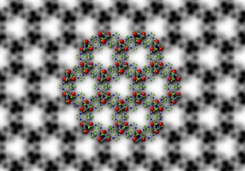
The team applied their method to ZIF-8, a MOF comprising zinc ions connected by organic 2-methylimidazole linkers. They were able to image its structure with a resolution of 0.21 nanometers (one nanometer is one billionth of a meter), a resolution high enough to image the individual columns of zinc atoms and organic linkers.

This helped the researchers to reveal the surface and interfacial structures of ZIF-8 crystals. "The results unraveled that porosity generated at the interfaces of ZIF-8 crystals is different from the intrinsic porosity of ZIF-8, which influences how gas molecules transport in ZIF-8 crystals," explained Han.

###

Media Contact

Michelle D'Antoni
[email protected]

http://kaust.edu.sa/

############

Story Source: Materials provided by Scienmag

Share13Tweet8Share2ShareShareShare2

Related Posts

Revolutionizing Blood Cancer Treatment: Reprogramming Cancer Cell Death to Activate the Immune System

October 10, 2025

LED Light Targets and Destroys Cancer Cells While Protecting Healthy Tissue

October 10, 2025

Upcoming Release: The Journal of Nuclear Medicine Ahead-of-Print Highlights – October 10, 2025

October 10, 2025

Wirth Named Fellow of the American Physical Society

October 10, 2025
Please login to join discussion

POPULAR NEWS

  • Sperm MicroRNAs: Crucial Mediators of Paternal Exercise Capacity Transmission

    1196 shares
    Share 478 Tweet 299
  • New Study Reveals the Science Behind Exercise and Weight Loss

    102 shares
    Share 41 Tweet 26
  • New Study Indicates Children’s Risk of Long COVID Could Double Following a Second Infection – The Lancet Infectious Diseases

    96 shares
    Share 38 Tweet 24
  • Revolutionizing Optimization: Deep Learning for Complex Systems

    83 shares
    Share 33 Tweet 21

About

We bring you the latest biotechnology news from best research centers and universities around the world. Check our website.

Follow us

Recent News

Revolutionizing Blood Cancer Treatment: Reprogramming Cancer Cell Death to Activate the Immune System

LED Light Targets and Destroys Cancer Cells While Protecting Healthy Tissue

Upcoming Release: The Journal of Nuclear Medicine Ahead-of-Print Highlights – October 10, 2025

Subscribe to Blog via Email

Enter your email address to subscribe to this blog and receive notifications of new posts by email.

Join 63 other subscribers
  • Contact Us

Bioengineer.org © Copyright 2023 All Rights Reserved.

Welcome Back!

Login to your account below

Forgotten Password?

Retrieve your password

Please enter your username or email address to reset your password.

Log In
No Result
View All Result
  • Homepages
    • Home Page 1
    • Home Page 2
  • News
  • National
  • Business
  • Health
  • Lifestyle
  • Science

Bioengineer.org © Copyright 2023 All Rights Reserved.