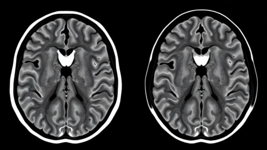
A stroke that silently threatens one side of the brain may announce itself in completely different ways depending on what is actually happening inside the artery wall. That is the central message of a new study published in Annals of Clinical and Translational Neurology, in which researchers used high-resolution magnetic resonance imaging to peer beneath the inner lining of narrowed brain arteries in nearly 400 adults with moyamoya vasculopathy. Their findings reveal that the imaging fingerprints of impending infarction are not universal: patients with classic moyamoya disease show one pattern of vascular failure, while those whose moyamoya-like changes are driven by atherosclerosis show an entirely different one. The discovery could reshape how clinicians decide which patients need surgery, and when.
Moyamoya vasculopathy is an umbrella term covering two related but distinct conditions. Moyamoya disease is a chronic, progressive narrowing of the terminal internal carotid arteries and the proximal segments of the anterior and middle cerebral arteries, accompanied by a hazy network of tiny collateral vessels at the base of the brain that resembles a puff of smoke on angiography, the meaning of the Japanese word moyamoya. Moyamoya syndrome, by contrast, produces a similar vascular picture but arises secondarily, most often when intracranial atherosclerosis mimics the appearance of the idiopathic disease. Distinguishing the two matters enormously, because their underlying biology, prognosis, and treatment strategies diverge, yet conventional angiography alone often cannot separate them reliably.
The research team, led by investigators at a major Chinese hospital registry, enrolled 398 consecutive adults with radiologically confirmed moyamoya vasculopathy between September 2017 and April 2025. Every patient underwent imaging on the same 3.0-Tesla MR scanner using a standardized protocol that included T1-weighted, T2-weighted, FLAIR, diffusion-weighted, and time-of-flight angiographic sequences, along with high-resolution vessel wall imaging. By scanning all participants on a single machine, the investigators eliminated the interscanner variability that often plagues multicenter imaging studies, allowing vessel wall measurements to be compared directly across the cohort.
High-resolution MRI has emerged in recent years as the decisive tool for separating the two etiologies. In moyamoya disease, the affected arteries typically show concentric, uniform wall thickening, a reduction in the outer diameter of the vessel, and negative or constrictive remodeling, meaning the artery wall shrinks inward as the disease progresses rather than expanding outward. Atherosclerosis-associated moyamoya vasculopathy displays the opposite signature: irregular, eccentric thickening caused by lipid-rich or hemorrhagic plaques, and positive or expansive remodeling, in which the vessel enlarges to accommodate plaque growth. Applying these criteria, the researchers classified 279 patients as having moyamoya disease and 61 as having the atherosclerosis-associated form.
The clinical profiles of the two groups differed in telling ways. Patients with moyamoya disease were more frequently female, consistent with the well-established female predominance of the idiopathic condition, and were significantly more likely to report a family history of early-onset stroke in first-degree relatives, a hallmark of the genetic predisposition linked to the RNF213 susceptibility gene. They also showed more frequent involvement of the posterior cerebral arteries, reflecting the diffuse, whole-circle-of-Willis nature of the disease. The atherosclerosis-associated group, though similar in age, lacked these familial and posterior-circulation signatures, reinforcing the view that it represents a secondary, plaque-driven process rather than a genetically driven arteriopathy.
To determine which imaging features actually tracked with stroke, the team took an unusually granular approach: they analyzed each cerebral hemisphere separately. Of the 558 hemispheres in the moyamoya disease group, 169 had suffered middle cerebral artery territory infarcts, visible as acute or chronic lesions on MRI, while 389 remained asymptomatic. Because the two hemispheres of the same patient are statistically correlated, the researchers used generalized estimating equations, a regression framework that treats each patient as a cluster, adjusting for age, sex, hypertension, diabetes, hyperlipidemia, smoking, alcohol use, and family history of early stroke. This design allowed each hemisphere to serve, in effect, as its own internal comparison.
The results in moyamoya disease were strikingly consistent. Ischemic hemispheres were independently associated with greater middle cerebral artery stenosis, more advanced Suzuki stage, and higher stenosis scores on the internal carotid, middle cerebral, and posterior cerebral arteries. The posterior cerebral artery score carried the strongest effect, with each one-point increase more than doubling the odds of ischemia. Intriguingly, the remodeling index of the middle cerebral artery showed the opposite relationship: hemispheres with more constrictive remodeling were more likely to be ischemic, suggesting that for a given degree of luminal narrowing, the inward shrinkage of the vessel wall compounds hemodynamic compromise, while better-preserved or compensating vessels maintain perfusion.
In the atherosclerosis-associated group, the picture inverted almost completely. Here, the degree of middle cerebral artery stenosis, the Suzuki stage, and the carotid and posterior circulation scores all lost their statistical significance after adjustment. What remained significant were the remodeling index of the middle cerebral artery, which was positively associated with ischemia, and involvement of the anterior cerebral artery. This pattern suggests that in plaque-driven moyamoya vasculopathy, it is not the raw percentage of narrowing that matters but how the vessel wall remodels around the plaque and whether the anterior circulation can still serve as a collateral pathway. Positive remodeling, long recognized as a marker of vulnerable atherosclerotic plaques, appears to signal danger rather than compensation in this group.
The authors are careful to frame these findings as cross-sectional associations rather than proven predictors of future stroke, noting that their definition of ischemic hemispheres included both acute and old infarcts, and that the retrospective, single-center design and the relatively small atherosclerotic subgroup impose limits on generalizability. They also acknowledge that a small fraction of patients lacked internal carotid artery involvement on angiography, a population that the revised 2022 diagnostic criteria would technically exclude, though sensitivity analyses excluding these patients left the results qualitatively unchanged. Even so, the hemisphere-level, cluster-adjusted analysis represents one of the most detailed noninvasive characterizations of moyamoya vasculopathy published to date.
The clinical implications are potentially far-reaching. For patients with moyamoya disease, quantitative measures such as stenosis severity, Suzuki stage, and combined arterial scores, together with evidence of constrictive remodeling, could help identify which hemisphere is at highest risk and guide the timing of revascularization surgery. For those with the atherosclerosis-associated form, attention should shift toward plaque behavior and anterior circulation collateral integrity rather than stenosis percentage alone, aligning management more closely with modern intracranial atherosclerosis care, where recent long-term evidence has questioned the value of stenting and pointed instead toward bypass options in selected cases. The study’s core lesson is that moyamoya vasculopathy is not one disease but two, and the artery wall itself, imaged at high resolution, tells clinicians which one they are treating.
Subject of Research: Etiology-specific associations between high-resolution MRI vessel wall features and cerebral infarction in adult moyamoya vasculopathy
Article Title: High‐Resolution MRI Revealed Different Etiology‐Specific Associations With Cerebral Infarction in Adult Moyamoya Vasculopathy
Article References: Han, G., Pan, J., Hong, Y., Fan, X., Yao, M., Zhou, L., Zhu, Y., Feng, F., & Ni, J. (2026). High‐Resolution MRI Revealed Different Etiology‐Specific Associations With Cerebral Infarction in Adult Moyamoya Vasculopathy. Annals of Clinical and Translational Neurology, 13(10), 2057-2067. https://doi.org/10.1002/acn3.70380
Image Credits: AI Generated
DOI: 10.1002/acn3.70380
Keywords: moyamoya disease, moyamoya vasculopathy, high-resolution MRI, vessel wall imaging, cerebral infarction, intracranial atherosclerosis, remodeling index, Suzuki stage, collateral circulation, stroke risk, RNF213, revascularization
News Source: Cassandra Pierce. (October 6, 2026). High-Resolution MRI Shows Stroke Risk Follows Different Rules in Moyamoya Disease and Its Atherosclerotic Mimic. Scienmag.