
In a remarkable advancement poised to refine cancer diagnostics, researchers have unveiled the potent combination of high-frame-rate contrast-enhanced ultrasound (HFR CEUS) with contrast vector imaging (CVI) for the evaluation of superficial lymph node (SLN) lesions. This cutting-edge approach promises more accurate discrimination between benign and malignant lymph nodes, potentially revolutionizing the clinical pathway for patients presenting with suspicious lymphadenopathy.
The innovative study, spearheaded by Li et al., was conducted at a single center between October 2023 and February 2024, enrolling 38 consecutive patients with suspected SLN anomalies. Employing a prospective methodology, the research team meticulously applied both conventional B-mode ultrasonography and the novel HFR CEUS combined with CVI post-processing to assess the diagnostic utility of these imaging modalities rigorously. The incorporation of fine-needle aspiration cytologic or histopathologic examination as a diagnostic gold standard underscored the robustness of their comparative analysis.
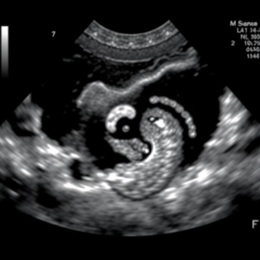
Central to the breakthrough is the technology of high-frame-rate contrast-enhanced ultrasound, which capitalizes on microbubble contrast agents and rapid image acquisition rates to visualize lymphatic perfusion dynamics with unprecedented temporal resolution. This technical enhancement enables clinicians to observe the microvascular flow patterns within lymph nodes in real time, capturing subtle differences that traditional ultrasound methods may miss.
Complementing this is contrast vector imaging, a sophisticated image processing technique that quantifies spatially and temporally resolved flow vectors within the enhanced ultrasound images. By translating intricate perfusion patterns into vectorial data, CVI offers a granular, quantitative perspective on lymph node vascularization, illuminating the pathophysiological underpinnings distinguishing malignant from benign nodes.
The study’s findings underscore a stark contrast in perfusion patterns between benign and malignant SLNs. Benign nodes predominantly exhibited centrifugal contrast dispersion, indicative of normal vascular architecture, whereas malignant nodes displayed centripetal and hybrid contrast patterns alongside frequent perfusion defects. These distinct vascular signatures, observable via HFR CEUS combined with CVI, provide a functional biomarker for malignancy with significant diagnostic implications.
Quantitatively, the synergy between HFR CEUS and CVI yielded a kappa coefficient of 0.81 when benchmarked against pathological diagnoses, denoting strong agreement. This surpasses the performance of HFR CEUS alone, which achieved a kappa value of 0.66, suggesting that the adjunctive use of CVI enhances diagnostic confidence and accuracy considerably.
The significance of these results is amplified by the study’s prospective design and the clinical relevance of superficial lymph nodes, which serve as accessible sentinel sites for metastatic spread in numerous cancers. Early and precise characterization of SLN lesions is critical for staging, treatment planning, and prognostication, positioning this imaging approach as an invaluable tool in oncologic care.
Technically, HFR CEUS leverages ultrafast imaging sequences that capture thousands of frames per second, surpassing conventional ultrasound frame rates by an order of magnitude. This advancement mitigates motion artifacts and enables a comprehensive temporal mapping of contrast agent kinetics within lymphoid tissue microcirculation, a feat unattainable with prior imaging protocols.
CVI’s contribution lies in its algorithmic capability to decompose and vectorize complex flow patterns, revealing directional blood flow trajectories and localized perfusion anomalies. This data-driven imaging modality enriches the interpretive framework beyond qualitative assessment, providing clinicians with objective metrics essential for nuanced differential diagnosis.
The integration of these technologies addresses long-standing limitations in lymph node evaluation where conventional B-mode ultrasound lacks sufficient specificity and contrast-enhanced ultrasound alone may fall short in characterizing heterogeneity within the nodal microenvironment. By merging rapid imaging acquisition with sophisticated computational analysis, this dual modality approach signifies a paradigm shift.
Beyond diagnostic superiority, the minimally invasive nature of HFR CEUS combined with CVI offers a patient-friendly alternative to more invasive procedures such as surgical biopsy. The ability to noninvasively monitor lymphatic changes in real time opens avenues for dynamic disease monitoring and personalized treatment adjustments, enhancing clinical outcomes.
However, the study also highlights the necessity for further large-scale validation to determine the reproducibility of these findings across diverse populations and varying clinical settings. Moreover, the standardization of imaging protocols and CVI parameter thresholds will be vital for widespread adoption and integration into routine diagnostic workflows.
The promising results reported herald a new era where real-time, high-resolution vascular imaging merges seamlessly with sophisticated computational tools to decode the complexities of lymph node pathology. This emergent technology stands to influence not only oncologic diagnostics but may also impact the evaluation of inflammatory and infectious lymphadenopathies, broadening its clinical utility.
In summary, the combined employment of high-frame-rate contrast-enhanced ultrasound and contrast vector imaging offers a potent, nuanced, and non-invasive approach to accurately differentiate benign from malignant superficial lymph node lesions. Its potential to enhance diagnostic precision, reduce reliance on invasive procedures, and tailor patient management marks a significant stride forward in medical imaging science.
As the oncology community seeks ever more refined diagnostic tools, the confluence of ultrafast imaging and advanced image processing demonstrated here represents a beacon of innovation. By illuminating the vascular signatures of malignancy with clarity and precision, this technology paves the way for more effective, timely, and personalized cancer care.
Such developments underscore the symbiotic relationship between technological ingenuity and clinical need, exemplifying how incremental scientific progress can culminate in transformative healthcare improvements. With ongoing research and clinical integration, HFR CEUS combined with CVI could soon become a mainstay in the diagnostic arsenal against cancer.
The study by Li et al. thus not only advances the frontier of lymph node imaging but also exemplifies the critical role of multidisciplinary collaboration in pushing the boundaries of diagnostic radiology. As technological capabilities evolve, their translation into clinically impactful solutions represents the hallmark of modern medical research.
Subject of Research: Diagnostic performance of high-frame-rate contrast-enhanced ultrasound combined with contrast vector imaging in detecting benign and malignant superficial lymph nodes.
Article Title: Diagnostic value of high-frame-rate contrast-enhanced ultrasound and contrast vector imaging for superficial lymph node lesions.
Article References:
Li, R., Lan, X., Xie, X. et al. Diagnostic value of high-frame-rate contrast-enhanced ultrasound and contrast vector imaging for superficial lymph node lesions. BMC Cancer 25, 785 (2025). https://doi.org/10.1186/s12885-025-14190-0
Image Credits: Scienmag.com
DOI: https://doi.org/10.1186/s12885-025-14190-0
Tags: benign vs malignant lymph nodescancer diagnostics technologycontrast vector imaging in ultrasoundfine-needle aspiration and histopathology in diagnosticshigh-frame-rate contrast-enhanced ultrasoundinnovative imaging techniques in medicinelymph node diagnosis advancementslymphadenopathy assessment methodsmicrobubble contrast agents in ultrasoundprospective study on ultrasound diagnosticsreal-time lymphatic perfusion imagingsuperficial lymph node evaluation


