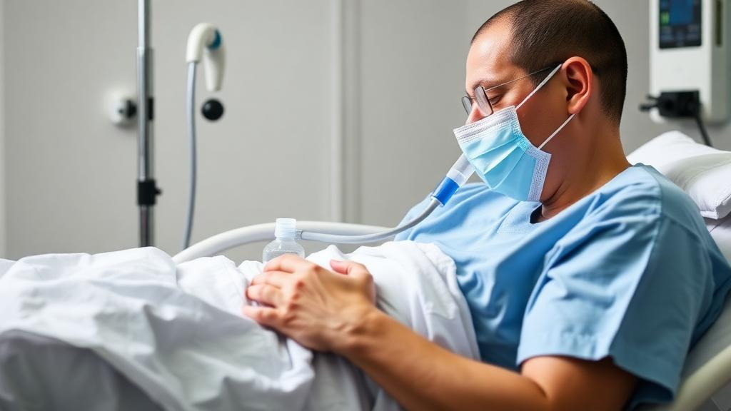
One of the most frustrating dilemmas in neurocritical care has long been the patient who can breathe but cannot yet protect their own airway. After brain tumor surgery, many people pass a spontaneous breathing trial with flying colors, only to remain tethered to a ventilator for days because impaired swallowing, weakened cough reflexes, or bulbar dysfunction make extubation unsafe. A new pilot study from Beijing Tiantan Hospital, published in Neurocritical Care, suggests that a simple technique—delivering warmed, humidified oxygen at high flow directly through the breathing tube—may allow these patients to break free from the ventilator while keeping the artificial airway in place as a safety net.
The clinical paradox the researchers set out to address is well known to intensive care physicians. In patients with structural brain injury, gas exchange and airway protection can become dissociated: the lungs and respiratory muscles may be perfectly capable of sustaining breathing, yet the neurological machinery guarding the airway lags behind. Conventional practice bridges this gap with low-level pressure support ventilation, in which the machine delivers small bursts of positive pressure with each breath. But that perceived safety may be deceptive. By offloading the inspiratory muscles, even modest pressure support can over-assist, suppressing the patient’s own respiratory drive and, over time, contributing to ventilator-induced diaphragmatic dysfunction—a condition that can transform a stable patient into one who is genuinely difficult to wean.
High-flow tracheal oxygen, or HFTO, offers a fundamentally different approach. Instead of cyclic pressure assistance, it delivers a continuous stream of warmed, humidified gas at high flow rates through the endotracheal tube. This mitigates the inspiratory resistance the tube itself imposes and provides a modest positive end-expiratory pressure effect, while leaving the work of breathing entirely to the patient. Evidence in tracheostomized patients has been emerging, but high-resolution physiological data in intubated patients had been scarce. The Beijing team, led by Yi-Min Zhou and Guang-Qiang Chen, designed a prospective randomized pilot study to fill that void, registering the trial on ClinicalTrials.gov and obtaining institutional ethics approval and informed consent from all participants or their legal representatives.
Between December 2025 and February 2026, the researchers screened thirty intubated patients within twenty-four hours of elective brain tumor resection. Twenty-four met the strict enrollment criteria: neurological stability with no intracranial hypertension for at least a day, a Glasgow Coma Scale score of at least nine, a successful thirty-minute spontaneous breathing trial on minimal support, and documented impairment of airway protection—assessed with a validated scoring tool—that was expected to persist for at least forty-eight hours. Patients with obesity, chronic obstructive pulmonary disease, asthma, or advanced heart failure were excluded. One patient was withdrawn after intracranial rebleeding, leaving twenty-three for analysis: twelve assigned to HFTO and eleven to low-level pressure support.
The study’s technical sophistication is what sets it apart. Rather than relying on clinical impressions, the team inserted esophageal manometry catheters, verified with the Baydur occlusion test, to measure the esophageal pressure–time product per minute—a gold-standard index of the work the inspiratory muscles perform with every breath. They also calculated the pressure generated by the inspiratory muscles and performed serial diaphragmatic ultrasound at baseline and at two, twelve, twenty-four, and forty-eight hours, tracking diaphragmatic thickness at end-expiration, the thickening fraction, and excursion during tidal breathing. Arterial blood gases, airway pressures, and hemodynamics were sampled at every time point, giving an unusually complete physiological portrait of what happens when positive pressure is withdrawn.
The primary finding was a striking divergence in the temporal trajectory of inspiratory effort. At baseline, the two groups were statistically indistinguishable. But repeated-measures analysis of variance revealed a significant group-by-time interaction, driven almost entirely by an abrupt physiological step-up in the HFTO group immediately after positive pressure support was withdrawn. Crucially, that elevated effort did not continue to climb; it stabilized at a safe plateau from two hours onward through the full forty-eight-hour observation window. The work of breathing did not progressively worsen or accumulate over time, which the authors note aligns with physiological expectations in patients with healthy lungs. The pattern for inspiratory muscle pressure mirrored the same trajectory.
Perhaps the most reassuring result concerned the diaphragm. Despite the higher workload, none of the ultrasound-derived indices—thickness, thickening fraction, or excursion—showed significant changes in either group, and no participant in either arm experienced a reduction in diaphragmatic thickness of twenty percent or more relative to baseline. This dissociation between increased global inspiratory effort and preserved diaphragmatic structure suggests that the loading imposed by HFTO remained within the adaptive capacity of the respiratory muscles over the short term. It also hints that HFTO may occupy a physiological sweet spot: enough activation to keep the muscle engaged, not so much as to cause injury.
Safety signals were largely favorable, with important nuances. Airway pressures in the HFTO group fell to near-zero or slightly negative values, in sharp contrast to the stable positive pressures in the ventilation group, and this difference was highly significant throughout. As expected with the loss of positive end-expiratory pressure, the ratio of arterial oxygen to inspired oxygen fraction declined modestly and carbon dioxide levels rose slightly in the HFTO group. Yet these changes did not translate into clinical failure: the rate of respiratory deterioration requiring escalation was statistically similar between groups, at twenty-five percent with HFTO versus 18.2 percent with pressure support. A minority of HFTO patients did exhibit episodes of inspiratory effort exceeding the predefined safety threshold, a reminder that removing both pressure assistance and end-expiratory pressure can unmask latent respiratory load in some individuals, and that the strategy demands careful monitoring rather than passive assumption of safety.
The clinical endpoints, though underpowered, offered an intriguing signal. Patients treated with HFTO accumulated a median of twenty-seven ventilator-free days at day twenty-eight, compared with twenty-one days in the pressure support group, a univariate difference the authors attribute primarily to earlier discontinuation of mechanical ventilation rather than earlier removal of the artificial airway itself. Artificial airway-free days and intensive care unit length of stay were comparable. Extubation success within seventy-two hours of study completion was similarly high in both arms, and reintubation and tracheostomy rates did not differ. No cases of inadequate humidification were reported, and no serious adverse events were attributable to the interventions.
The authors are careful to frame these findings as hypothesis-generating rather than practice-changing. The study was a single-center pilot with a small sample, an open-label design, and a narrowly defined population of relatively preserved brain tumor patients; the results cannot be extrapolated to severe traumatic brain injury, intracranial hemorrhage, or patients with obesity and lung disease. The design also cannot disentangle whether the observed effects stem from withdrawing cyclic pressure assistance, losing positive end-expiratory pressure, or both. Still, the conceptual contribution is substantial: liberation from the ventilator and removal of the artificial airway may be two distinct, separable steps in neurosurgical care. By acting as a kind of physiological stress test, high-flow tracheal oxygen could spare rapidly recovering patients unnecessary tracheostomies while identifying those with persistent deficits who truly need early surgical airways. Larger multicenter trials will now be needed to determine whether this elegant decoupling of ventilation from airway management delivers tangible benefits across the broader neurocritical population.
Subject of Research: Physiological comparison of high-flow tracheal oxygen and pressure support ventilation in neurosurgical patients with delayed extubation after brain tumor surgery
Article Title: Physiological Effects of High-Flow Tracheal Oxygen vs. Pressure Support Ventilation in Neurosurgical Patients with Brain Tumors Facing Delayed Extubation: A Pilot Study
Article References: Zhou, Y.-M., Ma, Y.-J., Tian, Y., Zhou, J.-F., Shi, G.-Z., Zhou, J.-X., & Chen, G.-Q. (2026). Physiological Effects of High-Flow Tracheal Oxygen vs. Pressure Support Ventilation in Neurosurgical Patients with Brain Tumors Facing Delayed Extubation: A Pilot Study. Neurocritical Care. https://doi.org/10.1007/s12028-026-02656-3
Image Credits: AI Generated
DOI: 10.1007/s12028-026-02656-3
Keywords: high-flow tracheal oxygen, pressure support ventilation, mechanical ventilation weaning, delayed extubation, neurocritical care, brain tumor surgery, diaphragmatic function, inspiratory effort, esophageal manometry, ventilator-induced diaphragmatic dysfunction, airway protection, pilot study
News Source: Ophelia Keating. (October 7, 2026). High-Flow Tracheal Oxygen Shows Promise for Brain Surgery Patients Stuck on Ventilators. Scienmag.