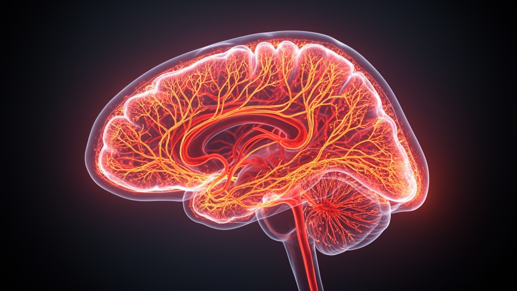
A high dose of methamphetamine rapidly elevates regional cerebral blood flow across multiple cortical and subcortical brain areas in anesthetized rats, producing a distinct forty-minute hemodynamic peak in the thalamus without altering large-vessel conductance blood flow in the Circle of Willis. The investigation, conducted by researchers at the Institute of Scientific Instruments of the Czech Academy of Sciences and Masaryk University in Brno, utilized high-field pharmacological magnetic resonance imaging to track how acute psychostimulant exposure modulates blood flow over a two-hour window. By tracking absolute perfusion levels using magnetically labeled arterial water, the researchers demonstrated that systemic administration of five milligrams per kilogram of methamphetamine causes immediate hyperperfusion in the whole brain, the sensorimotor cortex, the piriform cortex, and the thalamus. These quantitative findings provide a dose-resolved and time-resolved hemodynamic map of the neurovascular consequences that accompany acute stimulant challenges in a preclinical rodent model.
Methamphetamine acts as a potent indirect sympathomimetic agent that triggers massive extracellular release of catecholamines, including dopamine, noradrenaline, and serotonin, throughout the central nervous system. Previous neurochemical studies have established that systemic drug challenges can drive extracellular dopamine concentrations to several hundred percent of their basal values within minutes. While the molecular mechanisms of monoamine transporter reversal and clearance inhibition are well characterized, understanding how these neurochemical surges translate into functional microvascular changes in living tissue has remained technically challenging. Chronic exposure is known to elicit profound monoaminergic neurotoxicity, reductions in transporter density, and structural white and gray matter abnormalities that contribute to severe addiction and high relapse rates. However, therapeutic development has been continually hampered by a lack of non-invasive tools capable of dynamically tracking real-time functional brain alterations during acute drug administration.
To overcome the limitations of conventional hemodynamic imaging, the investigators turned to arterial spin labeling pharmacological magnetic resonance imaging. Although blood oxygen level-dependent imaging is widely deployed in functional neuroimaging, the resulting signal reflects an intertwined combination of cerebral blood flow, vascular volume, and cerebral metabolic rate of oxygen, making it difficult to isolate which physiological parameter changes during a pharmacological intervention. In contrast, arterial spin labeling employs magnetically inverted arterial blood water protons as an endogenous tracer, delivering quantitative, non-invasive measurements of regional cerebral blood flow in absolute units. Because regional perfusion is tightly coupled to local neuronal and glial activity through neurovascular coupling mechanisms mediated by vasoactive messengers, quantitative perfusion measurements serve as an indirect functional readout of drug actions across the neurovascular unit under controlled experimental conditions.
The experimental protocol evaluated ten male Sprague-Dawley rats that were nine weeks old upon arrival and between eleven and fourteen weeks old during imaging sessions. Animals were scanned under stable isoflurane inhalation anesthesia using an ultra-high-field 9.4-tesla Bruker small-animal magnetic resonance imaging system equipped with a volume transmitter coil and a four-channel rat head receiver array. To ensure rigorous comparisons across drug treatments while minimizing inter-subject variability, the team implemented a balanced Latin square crossover design. Over four consecutive scanning sessions separated by one-week washout intervals, every animal received an intraperitoneal injection of vehicle saline or methamphetamine at doses of one, two, or five milligrams per kilogram. These dosages were deliberately chosen to reflect behaviorally relevant self-administration thresholds established in previous operant conditioning literature, spanning low short-access intake up to extended-access binge consumption.
The team gathered perfusion measurements using a flow-sensitive alternating inversion recovery rapid acquisition with relaxation enhancement sequence with an acquisition time of eight minutes per run. After anatomical scout imaging and an initial basal perfusion assessment conducted thirty minutes after anesthetic induction, the animals received their designated systemic injections. The researchers subsequently acquired six consecutive arterial spin labeling scans at regular twenty-minute intervals, monitoring perfusion dynamics through one hundred twenty minutes post-injection while animal body temperature and respiration were continuously stabilized on a heated bed.
Perfusion maps calculated with specialized reconstruction software were inspected using blinded region-of-interest analysis referencing a standardized stereotaxic rat brain atlas. Evaluated structures included the whole-brain cross-section, the large conductance vessels comprising the Circle of Willis, and four bilateral parenchymal regions: the sensorimotor cortex representing neocortical sensorimotor networks, the piriform cortex representing paleocortical processing, the hippocampus involved in learning and contextual memory, and the thalamus acting as a central relay and arousal hub. Prior to grouping bilateral data, correlation analyses demonstrated very strong concordance between the right and left hemispheres across the sensorimotor cortex, hippocampus, and thalamus, justifying the use of averaged bilateral values for comprehensive statistical modeling using repeated-measures linear mixed-effects frameworks.
Initial baseline evaluations confirmed that the one-week clearance intervals effectively eliminated hemodynamic carry-over between experimental sessions, as basal blood flow values exhibited no significant differences among treatment groups across any evaluated brain region. Under basal conditions, absolute blood flow demonstrated a steep physiological gradient across anatomical territories. The Circle of Willis displayed the highest basal perfusion, registering approximately one hundred sixteen to one hundred twenty milliliters per minute per one hundred grams of tissue, reflecting its role as a low-resistance conductance arterial network. Among parenchymal areas, the thalamus exhibited substantially higher baseline perfusion, averaging between ninety-two and one hundred six milliliters per minute per one hundred grams, than the neocortical and paleocortical regions, which registered baseline values between sixty-five and seventy-eight milliliters per minute per one hundred grams.
Following pharmacological intervention, linear mixed-effects modeling revealed that methamphetamine exerts pronounced dose-dependent and region-specific effects on cerebral perfusion. The highest dose of five milligrams per kilogram elicited substantial hyperperfusion, elevating blood flow across the whole brain slice, the sensorimotor cortex, the piriform cortex, and the thalamus. In the sensorimotor cortex, perfusion surged by approximately thirty percent relative to baseline, remaining significantly higher than vehicle and lower drug doses. The piriform cortex exhibited a consistent increase of approximately fifteen percent. In stark contrast, neither the one milligram nor two milligrams per kilogram doses produced statistically significant hyperperfusion in these cortical structures, indicating a pharmacological threshold under isoflurane anesthesia below which monoamine release is insufficient to overcome anesthetic-induced basal vasodilation.
The thalamus demonstrated the most pronounced hemodynamic alteration observed in the entire investigation, displaying a dramatic sixty percent perfusion increase above baseline following the five milligrams per kilogram challenge. Furthermore, the thalamus was the only brain region to display a statistically significant temporal peak, reaching maximal hyperperfusion precisely forty minutes after drug injection before gradually declining back toward baseline values by one hundred twenty minutes. This temporal profile closely aligns with previous neurochemical microdialysis measurements documenting that extracellular dopamine overflow peaks between twenty and forty minutes following an identical systemic methamphetamine dose. The exceptional magnitude of the thalamic response likely reflects its dense capillary vascularity, intense metabolic demand, and convergent noradrenergic and serotonergic innervation from ascending brainstem projections that regulate sensory gating and arousal states.
Unlike the uniform hyperperfusion observed in cortical and thalamic structures, the hippocampus exhibited a divergent, non-monotonic response across drug doses. The authors noted that this complex dose-response profile may stem from opposing neurovascular signaling pathways linked to differential dopamine receptor subtype activation, wherein stimulating D1-like receptors promotes microvascular vasodilation whereas activating D2-like receptor families triggers vasoconstriction. However, interpretation remains cautious because the vehicle-treated control animals also exhibited a gradual twenty percent increase in hippocampal perfusion over the extended scanning period, suggesting potential regional interactions with isoflurane anesthesia.
Crucially, the large conductance vessels of the Circle of Willis showed no significant perfusion changes following methamphetamine administration at any dose or time point. This stability underscores the distinct physiological regulation governing cerebral conductance arteries compared to parenchymal microvascular beds. While parenchymal microvessels dilate in response to local neurovascular coupling signals generated by activated neurons and astrocytes releasing vasoactive messengers such as nitric oxide, dopamine, and neuropeptides, the Circle of Willis is primarily governed by systemic arterial blood pressure, myogenic autoregulation, and arterial carbon dioxide tensions. Consequently, localized surges in parenchymal monoamines do not alter gross blood inflow entering the base of the brain, localizing the drug effect directly to parenchymal microcirculation.
The authors contextualized their rodent observations against existing human and animal literature, noting both parallels and methodological distinctions. While prior rodent studies using amphetamine, apomorphine, and cocaine reported parenchymal hyperperfusion or elevated cerebral blood volume, human imaging studies in conscious volunteers have frequently documented cortical hypoperfusion following acute stimulant challenges. This divergence likely reflects methodological factors, including conscious versus anesthetized physiological states, peripheral cardiovascular counter-regulation, and species-specific differences in monoaminergic receptor distributions. Furthermore, clinical investigations in individuals with chronic methamphetamine use disorder during abstinence reveal widespread cerebral hypoperfusion, presenting a sharp contrast to acute intoxication and highlighting a multi-stage neuroimaging trajectory across stages of stimulant use and withdrawal pathology.
Several methodological boundaries circumscribe the generalizability of the findings and highlight avenues for future research. The study examined only male rats, precluding assessments of sex-dependent differences in neurovascular sensitivity despite established evidence that stimulant toxicity and behavioral responses vary by biological sex. Isoflurane anesthesia is known to uncouple blood flow from metabolism and elevate baseline perfusion, potentially dampening low-dose pharmacological responses. Because systemic blood pressure and arterial carbon dioxide were not continuously recorded, systemic cardiovascular contributions cannot be definitively excluded. Nonetheless, pulsed arterial spin labeling demonstrated high precision as a quantitative biomarker of stimulant pharmacodynamics, offering a validated imaging framework to evaluate future therapeutic interventions.
Subject of Research: Pharmacological arterial spin labeling MRI assessment of acute methamphetamine-induced regional cerebral blood flow changes in rats
Article Title: Acute methamphetamine produces dose- and time-dependent regional hyperperfusion in the rat brain: an arterial spin labeling phMRI study
Article References: Drazanova, E., Kratka, L., Harastova-Pavlova, I., & Ruda-Kucerova, J. (2026). Acute methamphetamine produces dose- and time-dependent regional hyperperfusion in the rat brain: an arterial spin labeling phMRI study. Frontiers in Pharmacology, 17, Article 2000280. https://doi.org/10.3389/fphar.2026.2000280
Image Credits: AI Generated
DOI: 10.3389/fphar.2026.2000280
Keywords: methamphetamine, arterial spin labeling, pharmacological MRI, regional cerebral blood flow, neurovascular coupling, thalamus, sensorimotor cortex, piriform cortex, hippocampus, Circle of Willis, Sprague-Dawley rats, dopaminergic signaling, hyperperfusion, cerebral hemodynamics, isoflurane anesthesia, FAIR-RARE sequence, small animal neuroimaging
News Source: Colin Clarke. (November 19, 2026). High-Dose Methamphetamine Triggers Acute Cerebral Hyperperfusion in Rat Brain Imaging Study. Scienmag.