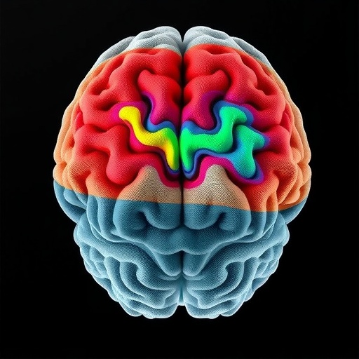
A new hierarchical map of the brain aims to unify rodent, primate, and human anatomy—turning multi-species brain scans into a single navigable atlas. In a study published in Nature Communications in 2026, researchers describe a computational framework that parcels both cortical and subcortical gray matter using a shared hierarchy across species, from rodents to primates and humans.
The core challenge is that brains differ in size, folding patterns, and landmark organization. Direct comparisons across species often break down because parcellations are typically optimized for a single anatomy. The new approach instead builds a multi-level structure that can align regions by their functional and structural relationships, rather than by surface coordinates alone.
To achieve this, the team integrates image-derived gray-matter features with hierarchical constraints. The model progressively groups small anatomical units into larger territories, while preserving consistency across scales. By doing so, it aims to reduce “one-off” region definitions and produce parcels that remain stable when transitioning between species.
Technically, the framework uses a data-driven segmentation strategy guided by an explicit hierarchy. Regions are not treated as isolated labels; they are organized as nested systems. This matters for subcortical structures, where boundaries can be subtle yet biologically important, and where misalignment can obscure cross-species correspondence.
The results suggest that the method can generate parcellations that are both species-specific enough to respect local anatomy and cross-species comparable enough to support translation. In other words, the atlas is designed to behave like a biological “dictionary,” where terms correspond to homologous architectural patterns rather than exact shapes.
Beyond mapping, the hierarchical design offers a practical advantage for neuroscience workflows. Researchers can map results from animal studies onto human coordinates more transparently, potentially improving how preclinical findings are interpreted during biomarker discovery or intervention planning.
The work also provides a foundation for building larger, community-accessible reference atlases. If adopted broadly, hierarchical parcellation could become a standard bridge between species, supporting consistent region definitions across laboratories and imaging platforms.
Viral science news aside, the study’s significance is simple: it tackles a long-standing bottleneck in comparative neuroanatomy by turning disparate brains into an interoperable framework. As cross-species datasets grow, this hierarchical atlas may become the backbone for next-generation comparisons of structure, development, and disease-related change.
Subject of Research: Multi-species cortical and subcortical gray-matter parcellation
Article Title: A hierarchical framework for cortical and subcortical gray-matter parcellation across rodents, primates, and humans
Article References: Venkadesh, S., Tian, Y., Linn, WJ. et al. A hierarchical framework for cortical and subcortical gray-matter parcellation across rodents, primates, and humans. Nat Commun (2026). https://doi.org/10.1038/s41467-026-75837-5
DOI: 10.1038/s41467-026-75837-5
Image Credits: AI Generated
Tags: brain structure alignment across speciescomputational brain atlas developmentcortical and subcortical brain segmentationcross-species neuroanatomical hierarchycross-species neuroanatomy comparisongray matter feature integrationhierarchical brain parcellation frameworkmulti-level brain mappingmulti-scale brain region organizationmulti-species brain atlasneuroimaging-based brain region delineationunified brain parcellation model