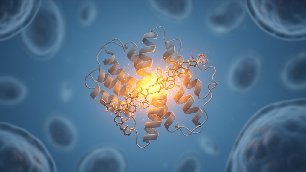
Scientists have long known that mucopolysaccharidosis type I, or MPS I, arises when a single lysosomal enzyme called α-ʟ-iduronidase (IDUA) stops doing its job. What they have not fully understood is how certain mutations leave the enzyme partially crippled rather than completely dead. Now, a team of Italian researchers has used extended molecular dynamics simulations to reveal a surprising answer: the damage may not lie in the protein’s backbone at all, but in the behavior of a sugar chain attached to it. The study, published in the Journal of Cellular and Molecular Medicine, identifies the conformation of the N372-linked glycan as a key molecular determinant of enzymatic impairment in attenuated MPS I.
MPS I is a rare autosomal recessive lysosomal storage disorder caused by mutations in the hIDUA gene. The enzyme belongs to glycosyl hydrolase family 39 and is responsible for cleaving α-linked ʟ-iduronic acid residues from glycosaminoglycans (GAGs), including dermatan sulphate and heparan sulphate. When IDUA is deficient, these GAGs accumulate inside lysosomes, causing impaired cellular function and multisystem damage. Clinical severity ranges from severe Hurler syndrome, characterized by early-onset neurodegeneration and skeletal abnormalities, to attenuated Scheie syndrome, which presents later in life with milder manifestations such as joint stiffness, corneal clouding, and cardiac involvement.
The researchers focused on two pathogenic variants, L490P and P533R, which are commonly associated with attenuated and intermediate phenotypes. These mutations retain partial IDUA activity, reducing systemic disease severity, but can still lead to mild neurological impairments due to GAG accumulation in the central nervous system. Current treatments, including enzyme replacement therapy and haematopoietic stem cell transplantation, can ameliorate somatic symptoms but are less effective in addressing CNS involvement because enzymes struggle to cross the blood–brain barrier. Emerging strategies such as gene therapy remain costly and require further validation.
To investigate the molecular mechanisms underlying mutation-induced dysfunction, the team prepared three-dimensional models of both wild-type and mutant IDUA using the experimentally solved X-ray structure (PDB code 4KGL). They ran molecular dynamics simulations at two different pH conditions: pH 7.5 to reflect the endoplasmic reticulum microenvironment, where protein folding and maturation occur, and pH 4.5 to mimic the lysosomal environment, where IDUA exerts its biological function. Production simulations totaled 1.6 microseconds across three independent replicas for each system, a substantial improvement over previous computational studies that were limited by shorter simulation times and did not account for distinct pH environments.
The results were striking. Backbone RMSD analysis showed that both wild-type and mutant proteins remain stable at both pH conditions, with no significant differences detected in the β-sandwich domain where both mutated residues are located. Secondary-structure analysis confirmed that the mutations do not substantially affect the protein’s overall fold within the simulated timescale. This finding challenges earlier suggestions that these mutations destabilize the local backbone of the β-sandwich domain and the neighbouring α15-helix.
Instead, the damage emerged when the researchers turned their attention to the N372-linked glycan. This glycan, composed of five saccharides—three mannose and two N-acetylglucosamine residues—is functionally unique among the six glycosylation sites on IDUA. Previous work by Maita and colleagues showed that it can directly interact with the substrate and that its occupancy positively correlates with enzymatic activity. At lysosomal pH, the wild-type protein displayed multiple glycan conformational states and higher flexibility, whereas both mutants exhibited a more restricted conformational behavior. RMSF values at pH 4.5 were markedly higher for the wild type (6.438 ± 1.641 Å SEM) compared to L490P (3.456 ± 0.850 Å SEM) and P533R (3.531 ± 0.204 Å SEM), indicating that mutations at positions 490 and 533 induce glycan rigidity.
Principal component analysis and clustering further revealed that the mutants occupy distinct conformational spaces compared to the wild type. Hydrogen-bond analysis provided mechanistic insight: in the L490P mutant, the glycan forms interactions with catalytic residue E182 and D187 that are practically absent in the wild type. The interaction with E182, one of the two key catalytic residues, provides a plausible hypothesis for impaired enzymatic activity, as the glycan’s repositioning toward the active-site region could influence substrate access. In the P533R mutant, a distinct interaction pattern emerged, characterized by a hydrogen bond between the glycan and residue D484 with significantly higher occupancy (29.70% for MAN6 and 17.42% for MAN7). D484 is located in proximity to the mutated site, suggesting that enhanced interaction with the glycan may arise from local structural rearrangements induced by the mutation.
Solvent-accessible surface area analysis of the binding pocket confirmed that the wild-type enzyme is more accessible compared to the mutants. To validate these computational predictions, the team performed preliminary in vitro measurements using a COS7 cell expression system. The residual enzymatic activity was 55% for L490P and 57% for P533R compared to the wild type, with both mutants differing significantly from the wild type (p < 0.0001). These comparable functional effects support the hypothesis that distinct structural perturbations converge on a similar functional outcome: restriction of glycan dynamics at lysosomal pH.
The study proposes a model in which disease-associated mutations impair IDUA function not by disrupting the global protein fold, but by restricting the conformational freedom and orientation of the N372-linked glycan. This glycan is not merely a passive post-translational modification but may act as an active conformational element whose flexibility and orientation are tightly coupled to enzymatic competence. Its perturbation by distal mutations may contribute to the residual but clinically significant enzymatic dysfunction characteristic of the Scheie phenotype.
These findings contribute to a more comprehensive understanding of MPS I pathogenesis and may provide a foundation for the rational design of therapeutic strategies aimed at modulating glycan–protein interactions or restoring productive glycan conformational dynamics—an aspect that remains largely underexplored. As the molecular details of how a sugar chain can act as a functional switch come into sharper focus, the possibility of targeting these interactions pharmacologically opens a new avenue for treating attenuated MPS I and potentially other lysosomal storage disorders where glycans play a similar regulatory role.
Subject of Research: Molecular Dynamics Reveals a Glycan Conformation Underlying IDUA Dysfunction in Attenuated MPS I
Article Title: Molecular Dynamics Reveals a Glycan Conformation Underlying IDUA Dysfunction in Attenuated MPS I
Article References: Papadopoulos, M. G. E., Bonapace, G., Taglietti, L., Ferla, B. L., Rizzi, R., Pitaro, M., Concolino, D., Alberga, D., & Mangiatordi, G. F. (2026). Molecular Dynamics Reveals a Glycan Conformation Underlying IDUA Dysfunction in Attenuated MPS I. Journal of Cellular and Molecular Medicine, 30(19), Article e71396. https://doi.org/10.1111/jcmm.71396
Image Credits: AI Generated
DOI: 10.1111/jcmm.71396
Keywords: MPS I, IDUA, lysosomal storage disorder, molecular dynamics, N372-linked glycan, L490P, P533R, glycan conformation, enzyme dysfunction, attenuated phenotype, Molecular, Dynamics
News Source: Drew Townsend. (October 17, 2026). Hidden Sugar Switch Sabotages a Key Enzyme in Attenuated MPS I. Scienmag.