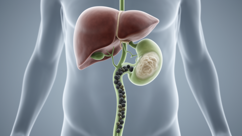
For more than a year, a 63-year-old woman in Guizhou, China, endured intermittent pain in her right upper abdomen. Her symptoms had been attributed to gallstones at another hospital, where symptomatic treatment brought only temporary relief. When the pain worsened over ten days, she arrived at the People’s Hospital of Anshun City with a history that seemed to point squarely at a common problem: stones in the biliary system causing recurrent discomfort. What her surgical team ultimately found inside her bile duct, however, was something far rarer and far more consequential—a precancerous tumor that had been hiding in plain sight behind a wall of stones and obstructed plumbing.
The case, published in Frontiers in Medicine by a team led by Jiankai Long, Jingnan Xue, and Kai Zhou of the People’s Hospital of Anshun City, with collaborators from Peking Union Medical College Hospital, describes an intrahepatic intraductal papillary neoplasm of the bile duct, or IPNB, with low-grade dysplasia of the pancreatobiliary type. IPNB is an uncommon biliary epithelial neoplasm characterized by papillary or villous proliferation within the bile duct lumen, often accompanied by bile duct dilatation and mucin hypersecretion. It is increasingly recognized as an important precursor lesion of cholangiocarcinoma, an aggressive cancer of the bile ducts. The histological grade, invasive status, and subtype of IPNB are closely associated with prognosis and management strategies, which makes accurate preoperative recognition a matter of genuine clinical urgency.
The diagnostic challenge in this case was not simply the rarity of the tumor itself. It was the fact that everything about the patient’s presentation—recurrent biliary symptoms, multiple intrahepatic stones, and evidence of ductal obstruction—could be explained by uncomplicated hepatolithiasis with secondary obstruction. In such settings, the main clinical risk is misinterpreting a true intraductal neoplasm as stone-related biliary disease. The authors emphasize that focal or unilateral bile duct dilatation, localized obstructive change, intraductal soft-tissue components, or abundant mucin may indicate IPNB rather than hepatolithiasis alone, and that recognizing this pitfall is essential for timely intervention.
On admission, physical examination revealed no remarkable findings. Laboratory investigations, including complete blood count, liver and renal function tests, and coagulation profile, were within normal limits. The only abnormality was a mildly elevated serum carbohydrate antigen 19-9, or CA19-9, level of 39.8 U/mL—a tumor marker that can be elevated in biliary malignancies but is nonspecific and can also rise in benign cholestatic conditions. Routine blood work offered no clear signal that a neoplasm was lurking within the left intrahepatic biliary system.
The real clues emerged from cross-sectional imaging. Contrast-enhanced computed tomography demonstrated a focal intraductal adenomatous lesion in the left intrahepatic biliary system with adjacent cystic or aneurysmal dilatation of the affected bile duct, producing the appearance of localized high biliary obstruction. The lesion was centered near the left hepatic hilar region and was associated with unilateral left intrahepatic biliary dilatation. Magnetic resonance imaging and magnetic resonance cholangiopancreatography, or MRCP, further confirmed marked unilateral dilatation of the left intrahepatic bile ducts with focal hilar-level obstructive change and an associated intraductal abnormality. The predominantly unilateral distribution of ductal dilatation and the localized obstructive appearance were atypical for simple hepatolithiasis alone and raised concern for an underlying intraductal biliary neoplasm.
This imaging pattern is technically significant because hepatolithiasis typically produces diffuse, bilateral, or multifocal ductal changes proportional to the stone burden. When dilatation is focal or unilateral and appears disproportionate to the stones, the differential diagnosis should expand beyond stone disease. In this case, the preoperative differential diagnosis included hepatolithiasis with cholangitis, intrahepatic cholangiocarcinoma, mucinous biliary neoplasm, and IPNB. The radiologic-pathologic correlation proved particularly informative: the focal intraductal lesion and adjacent dilated bile duct seen on CT corresponded to the gross finding of papillary adenomatous tissue within a markedly dilated bile duct containing gelatinous material, and to the microscopic finding of papillary epithelial proliferation on histopathology. This correlation helped explain why the lesion was not merely a consequence of stone disease but represented a true intraductal neoplasm.
Given that the lesion appeared localized to the left intrahepatic biliary system and that the patient had recurrent obstructive symptoms, the surgical team selected definitive treatment after comprehensive preoperative evaluation. The patient underwent open left hepatectomy under general anesthesia. Intermittent inflow occlusion using the Pringle maneuver—a technique that temporarily clamps the hepatoduodenal ligament to reduce blood loss during liver resection—was applied, with a total clamping time of 30 minutes. Intraoperative blood loss was approximately 20 mL, and the patient remained hemodynamically stable throughout the procedure. Gross examination of the resected specimen demonstrated a markedly dilated bile duct containing intraductal papillary adenomatous tissue and gelatinous mucinous material. For lesions confined to one intrahepatic biliary system, hepatectomy can provide both definitive diagnosis and curative treatment in a single operation.
Histopathological examination confirmed intrahepatic IPNB with low-grade dysplasia of the pancreatobiliary type. No invasive carcinoma was identified, and all resection margins were negative—findings that carry substantial prognostic weight. Immunohistochemical staining showed that the neoplastic biliary epithelium was positive for CK19, CK7, MUC1, MUC5AC, and MUC6, but negative for MUC2. This mucin profile is technically meaningful because it supports pancreatobiliary differentiation and strengthens subtype classification beyond morphology alone, enabling more meaningful comparison with previously reported cases. The postoperative course was uneventful, and the patient was discharged on postoperative day 10 without documented complications such as bile leakage, hemorrhage, or severe infection.
During two years of outpatient follow-up, the patient remained clinically stable with no evidence of recurrence or major postoperative complications. Her abdominal symptoms resolved after surgery, and her quality of life improved. The latest CT demonstrated postoperative changes after left hepatectomy, possible intrahepatic bile duct calculi or calcifications, mild intrahepatic and extrahepatic bile duct dilatation, and a right renal cyst, but no definite recurrent lesion was reported. After surgery, the patient reported that her pain had resolved and that follow-up examinations were reassuring, expressing satisfaction with the treatment outcome and feeling more secure in her daily life.
The authors caution that even low-grade, completely resected IPNB requires long-term surveillance, because biliary neoplasms may recur or present as multifocal disease. Patients with IPNB should undergo periodic clinical review and hepatobiliary imaging after surgery, especially during the first two postoperative years. The report has limitations: several detailed preoperative radiologic parameters, including reproducible lesion measurements, formal enhancement characteristics, and complete archived MRCP descriptors, were unavailable, and as a single-case report the generalizability of the findings is inherently limited. The two-year follow-up duration may also be insufficient to exclude late recurrence or multifocal disease. Nevertheless, the central message stands: in patients with hepatolithiasis and recurrent biliary symptoms, unilateral or focal intrahepatic duct dilatation, localized obstructive change, and intraductal abnormalities should prompt consideration of IPNB rather than uncomplicated stone disease alone. Early recognition, timely surgical resection for localized disease, and complete pathological evaluation are essential for accurate diagnosis, risk stratification, and follow-up planning—and this case shows how a tumor hiding behind stones can be caught before it becomes cancer.
Subject of Research: Medicine
Article Title: Intrahepatic intraductal papillary neoplasm of the bile duct with low-grade dysplasia presenting as high biliary obstruction and hepatolithiasis: a case report
Article References: Long, J., Xue, J., Xie, N., Zhang, L., & Zhou, K. (2026). Intrahepatic intraductal papillary neoplasm of the bile duct with low-grade dysplasia presenting as high biliary obstruction and hepatolithiasis: a case report. Frontiers in Medicine, 13, Article 1916026. https://doi.org/10.3389/fmed.2026.1916026
Image Credits: AI Generated
DOI: 10.3389/fmed.2026.1916026
Keywords: IPNB, bile duct, hepatolithiasis, cholangiocarcinoma, case report, hepatectomy, biliary obstruction, precancerous, mucin, diagnostic pitfall, Intrahepatic, intraductal
News Source: Ophelia Keating. (October 18, 2026). Hidden Bile Duct Tumor Masquerades as Gallstones in Landmark Case Report. Scienmag.