Researchers from Heilongjiang University of Chinese Medicine and affiliated hospitals reported that Xiebai Bining Decoction (XBD) reduced renal fibrosis and improved kidney function in a mouse model of hyperuricemic nephropathy. The study, published in Frontiers in Cell and Developmental Biology, demonstrated that the traditional Chinese medicine formulation was associated with lower serum uric acid, creatinine, and blood urea nitrogen levels compared to a model group. These findings suggest that XBD may offer renal protection that extends beyond simple urate lowering, although the study was conducted exclusively in mice and did not establish a direct causal mechanism between the observed molecular changes.
Hyperuricemic nephropathy is a significant renal complication of hyperuricemia, characterized by renal tubular injury and progressive tubulointerstitial fibrosis. The condition can eventually lead to renal failure, with sustained elevation of uric acid potentially aggravating tubular injury through oxidative stress, mitochondrial dysfunction, and inflammatory responses. The transforming growth factor-β1 (TGF-β1)/Smad3 signaling pathway is a well-established profibrotic pathway that promotes epithelial-mesenchymal transition and excessive extracellular matrix deposition. Additionally, mitochondrial dysfunction is increasingly recognized as an important contributor to renal fibrosis, making mitochondrial quality control a relevant area of investigation.
The research team utilized a potassium oxonate- and adenine-induced hyperuricemic nephropathy mouse model to evaluate the effects of XBD. Male Institute of Cancer Research (ICR) mice were randomly divided into six groups, including a blank control, a model group, and groups treated with febuxostat, XBD, the autophagy inhibitor 3-methyladenine (3-MA), or a combination of XBD and 3-MA. The XBD formulation consisted of five herbs: stir-fried Cangzhu, stir-fried Huangbai, Chuanniuxi, Bixie, and Tufuling. The decoction was prepared by traditional methods and administered via oral gavage at a dose of 520 mg/kg/day for 17 days, a dose corresponding to approximately 42.2 mg/kg/day in humans based on body-surface-area scaling.
Renal function was assessed by measuring serum uric acid, creatinine, and blood urea nitrogen levels. Histopathological evaluations included hematoxylin and eosin staining for general tissue morphology and Masson’s trichrome staining to quantify collagen deposition in the renal interstitium. The researchers also examined molecular markers of fibrosis and autophagy using Western blotting, immunofluorescence staining, immunohistochemistry, enzyme-linked immunosorbent assay, and transmission electron microscopy. These methods allowed for a comprehensive assessment of both structural and molecular changes in the kidney tissue.
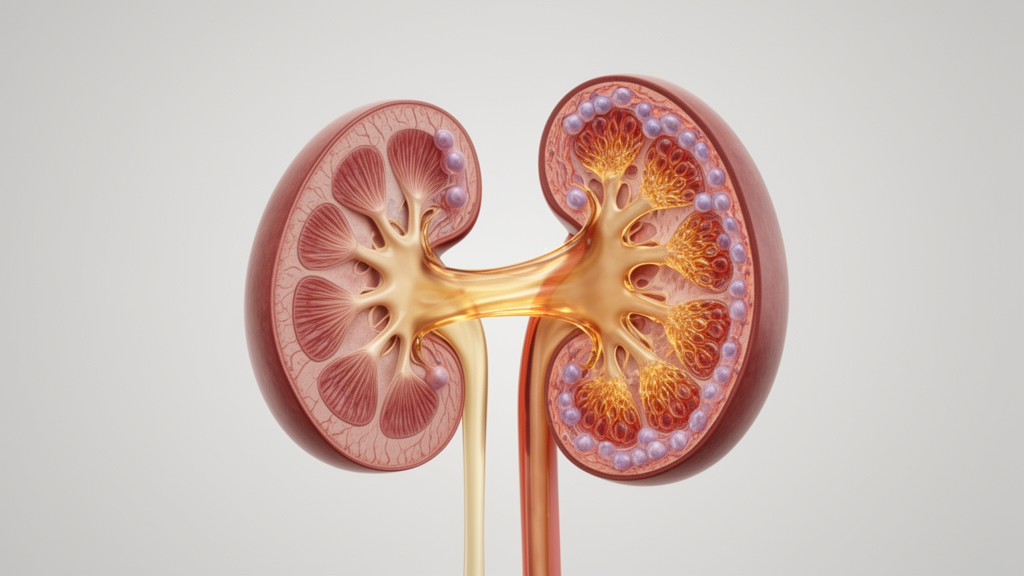
The results showed that XBD treatment significantly reduced serum uric acid, creatinine, and blood urea nitrogen levels compared to the untreated model group. XBD also improved gross kidney morphology, reducing the enlarged volume and yellowish-white deposits observed in the model mice. Histological analysis revealed that XBD alleviated renal tubular injury, including tubular dilation, epithelial cell edema, and vacuolar degeneration. Notably, the improvements in renal function and morphology with XBD were superior to those observed with febuxostat, a standard xanthine oxidase inhibitor used as a positive control in the study.
Regarding renal fibrosis, Masson’s trichrome staining indicated that XBD intervention markedly inhibited collagen deposition in the renal interstitium. Western blot analysis confirmed that XBD downregulated the expression of profibrotic proteins, including Collagen I, matrix metalloproteinase-9, and α-smooth muscle actin, while upregulating the epithelial marker E-cadherin. Immunohistochemical staining further supported these findings, showing reduced deposition of Collagen I and MMP-9 in the renal interstitium of XBD-treated mice. These changes suggest an attenuation of the fibrotic tissue response and preservation of epithelial markers.
The study also investigated the role of autophagy and mitochondrial quality control. Transmission electron microscopy revealed that XBD treatment improved mitochondrial ultrastructure and significantly increased the number of cytosolic autophagosomal profiles compared to the model group. Western blotting and immunofluorescence staining showed higher expression levels of FUN14 domain-containing protein 1 (FUNDC1) and microtubule-associated protein 1 light chain 3B (LC3B) in the XBD group. FUNDC1 is a mitophagy receptor located on the outer mitochondrial membrane, while LC3B is associated with autophagosomal membranes. The increase in these markers suggests an association with an autophagy-associated mitochondrial response.
Furthermore, XBD treatment was associated with changes in the TGF-β1/Smad3 signaling pathway. ELISA results showed that XBD reduced TGF-β1 content in renal tissue, while Western blotting indicated lower expression of TGF-β receptor type I and phosphorylated Smad3. These molecular changes occurred alongside reduced interstitial collagen deposition and fewer α-SMA-positive cells. The researchers noted that while these findings link lower TGF-β1/Smad3-associated readouts with a less fibrotic renal phenotype, they do not establish a direct regulatory interaction between the autophagy markers and the fibrosis signaling pathway.
The use of 3-methyladenine, an autophagy inhibitor, provided additional insights into the relationship between autophagy and renal injury. Mice treated with 3-MA alone exhibited more pronounced mitochondrial damage, fewer autophagosomal profiles, and exacerbated renal function deterioration compared to the model group. However, co-administration of XBD with 3-MA improved several outcomes relative to 3-MA alone, including renal morphology and molecular marker expression. This suggests that the renal protective effects of XBD may be partially mediated through autophagy-related processes, although 3-MA is not a FUNDC1-selective intervention and cannot establish the specific contribution of this mitophagy receptor.
The study has several limitations that must be considered when interpreting the results. The mouse model relies on potassium oxonate to inhibit uricase, a enzyme that is functional in mice but not in humans, and adenine exposure can contribute to tubular injury independently of hyperuricemia. The study used only male mice, so potential sex-related differences in renal urate handling and treatment responses remain to be investigated. Additionally, the chemical composition of the XBD decoction was not fully resolved, and the observed renal responses cannot be assigned to specific compounds or component-target interactions. Future studies will need to include chemical profiling of the finished decoction and controlled clinical trials to evaluate safety and efficacy in humans.
The findings contribute to the growing body of evidence regarding the potential of traditional Chinese medicine in managing hyperuricemic nephropathy. While previous studies have shown that various herbal formulations can influence urate metabolism and inflammatory responses, this study provides a detailed mechanistic exploration of XBD in the context of renal fibrosis and mitochondrial quality control. The concurrent changes in FUNDC1/LC3B markers and TGF-β1/Smad3 signaling readouts identify parallel molecular responses that warrant further targeted mechanistic validation. The research highlights the complexity of translating pathway modulation into clinical benefit and underscores the need for rigorous preclinical and clinical evaluation.
In conclusion, Xiebai Bining Decoction ameliorated renal dysfunction, pathological injury, and renal fibrosis in hyperuricemic nephropathy mice. These benefits were accompanied by changes in autophagy markers and lower TGF-β1/Smad3-related measurements, although a causal relationship between these pathways was not established. The study supports the potential of XBD as an intervention for HN-associated renal fibrosis but emphasizes the necessity for further investigation to define the optimal dose, chemical basis, and clinical applicability of the formulation.
The formula’s composition is rooted in Ermiao San from Danxi Xinfa, retaining fifteen grams each of stir-fried Cangzhu and stir-fried Huangbai to clear heat and dry dampness. To complete the prescription, the formulation incorporates fifteen grams of Chuanniuxi to promote blood circulation and guide treatment downward, ten grams of Bixie to resolve dampness and separate the clear from the turbid, and ten grams of Tufuling to dispel dampness and resolve toxin-related patterns. The preparation followed traditional aqueous extraction by soaking the combined botanicals in eight volumes of water for thirty minutes, boiling the mixture for one hour, repeating the extraction once, and filtering the combined batches before concentration and lyophilization into powder.
The testing protocol implemented a staged schedule across sixty male ICR mice that were eight weeks old and weighed twenty to twenty-three grams. Experimental groups of ten animals began with a three-day preventive phase where oral XBD, oral febuxostat at five milligrams per kilogram daily, or intraperitoneal 3-MA at fifteen milligrams per kilogram daily was administered at eight-thirty in the morning. Disease induction commenced on Day four via daily intraperitoneal injections of potassium oxonate at 350 milligrams per kilogram alongside intragastric adenine at seventy milligrams per kilogram, continuing through Day seventeen. While ten mice were assigned to each experimental group for baseline evaluations, renal tissue samples from three randomly selected mice per cohort were specifically allocated for core molecular assays, including Western blotting, transmission electron microscopy, and ELISA.
Additional molecular examinations highlighted changes in phosphorylated Histone H3, a marker whose elevated expression in model mice was reversed by XBD. Kidney weight and the kidney coefficient, calculated as kidney mass divided by body weight, were similarly elevated in untreated model mice and normalized by decoction therapy. However, the authors emphasized that static autophagosomal profiles on electron microscopy and elevated FUNDC1 abundance do not directly quantify mitophagic flux or prove cargo clearance. Because 3-MA is not a selective intervention, it cannot confirm a receptor-dependent role for FUNDC1.
The investigation was also restricted to a single fixed dosage of 520 milligrams per kilogram daily, meaning that an optimal dosing range or dose-response relationship was not established. In addition, while individual constituent herbs contain recognized compounds such as atractylodin in Cangzhu, berberine in Huangbai, and astilbin in Tufuling, the administered decoction was not characterized through liquid chromatography-tandem mass spectrometry. Consequently, specific bioactive molecules responsible for the observed renal effects remain unverified until comprehensive chemical profiling and subsequent target validation are conducted.
Subject of Research: Xiebai bining decoction attenuates renal fibrosis in hyperuricemic nephropathy alongside changes in FUNDC1/LC3B autophagy markers and TGF-β1/Smad3 signaling
Article Title: Xiebai bining decoction attenuates renal fibrosis in hyperuricemic nephropathy alongside changes in FUNDC1/LC3B autophagy markers and TGF-β1/Smad3 signaling
Article References: Li, K., Xia, X., Li, M., & Tong, Y. (2026). Xiebai bining decoction attenuates renal fibrosis in hyperuricemic nephropathy alongside changes in FUNDC1/LC3B autophagy markers and TGF-β1/Smad3 signaling. Frontiers in Cell and Developmental Biology, 14, Article 1975823. https://doi.org/10.3389/fcell.2026.1975823
Image Credits: AI Generated
DOI: 10.3389/fcell.2026.1975823
Keywords: Xiebai, bining, decoction, attenuates, renal, fibrosis, hyperuricemic, nephropathy, alongside, changes, FUNDC1, LC3B, autophagy, markers, TGF-
News Source: Drew Townsend. (November 14, 2026). Herbal Decoction Reduces Kidney Fibrosis in Hyperuricemic Mouse Model. Scienmag.