


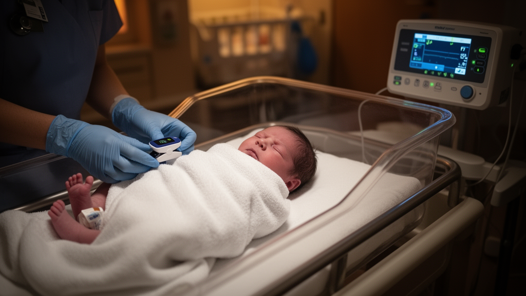
Infants born with congenital cyanotic heart disease may carry hidden auditory risks long before they ever reach an operating table. A new cross-sectional study from Tanta University Hospital in Egypt reports that nearly half of a group of young infants with cyanotic heart defects showed elevated auditory brainstem response thresholds on a single preoperative hearing test, with most abnormalities mild and many affecting only one ear. The findings, published in the Egyptian Pediatric Association Gazette, add to growing evidence that the developing auditory system may be vulnerable in children whose blood oxygen levels run chronically low.
The research team enrolled 51 infants aged between 6 and 24 months who had been diagnosed with congenital cyanotic heart disease and had not yet undergone any corrective or palliative cardiac surgery. All participants were recruited consecutively from the Pediatric Cardiology Unit between December 2025 and May 2026. Of 62 infants screened, eleven were excluded for reasons including prior cardiac surgery, recurrent otitis media, syndromic diagnoses associated with hearing loss, or incomplete audiological data. The final cohort had a mean age of 14.9 months, and just over half of the infants, 26 of 51, were female. Most families came from rural areas, and 17 infants, roughly one third, had a history of neonatal intensive care unit admission.
The cardiac diagnoses reflected the typical spectrum of cyanotic defects. Tetralogy of Fallot was the most common condition, affecting 19 infants or 37.3 percent of the cohort, followed by transposition of the great arteries in 17 infants, or 33.3 percent. Double outlet right ventricle accounted for 19.6 percent and tricuspid atresia for 9.8 percent. The mean baseline oxygen saturation recorded at assessment was 80.61 percent, a figure that underscores how profoundly these infants live below normal blood oxygen levels. The authors stress, however, that this value reflects oxygenation at the moment of testing rather than a standardized measure of long-term hypoxemic exposure, because some infants were assessed while receiving their prescribed supplemental oxygen.
Each infant underwent a structured audiological battery before any surgical intervention. Otoscopy excluded outer and middle ear pathology, and tympanometry with a 226 Hz probe tone assessed middle ear function. Auditory brainstem response testing was then carried out during natural quiet sleep, without sedation, in an electrically shielded room using click stimuli delivered through headphones. Testing began at 90 decibels normalized hearing level and descended in 10-decibel steps, then 5-decibel steps near threshold, until the lowest intensity producing a repeatable wave V response was identified. A wave V threshold at or below 30 dBnHL was considered normal, while severity was graded as mild, between 31 and 40 dBnHL, or moderate-to-severe, at 41 dBnHL and above, following a World Health Organization classification adapted for infant ABR testing.
The results were striking in their pattern. Twenty-two infants, or 43.1 percent, showed click-ABR wave V threshold elevations above 30 dBnHL on this single assessment. Ten of these, 19.6 percent, had bilateral elevations, while twelve, 23.5 percent, had elevations in only one ear. At the ear level, among the 102 ears examined, 18 left ears and 14 right ears showed elevated thresholds. Most affected ears fell in the mild range: 14 of the 18 affected left ears and 10 of the 14 affected right ears had thresholds of 31 to 40 dBnHL. Only four infants, 7.8 percent of the cohort, demonstrated moderate-to-severe bilateral elevations, with two showing thresholds of 60 dBnHL and two showing 70 dBnHL bilaterally. Both infants with severe elevations had histories of neonatal intensive care admission.
Every ear with an elevated ABR threshold showed a Type A tympanogram, indicating no overt middle-ear dysfunction at the time of testing and making a conductive component less likely. Yet the authors are careful about what these findings do and do not establish. Because click-evoked ABR combined with normal tympanometry cannot definitively distinguish sensorineural hearing loss from other types of impairment, and because no repeat testing or longitudinal follow-up was systematically performed, the 43.1 percent figure represents single-assessment threshold elevations rather than confirmed permanent hearing loss. The team also notes that some of the mild elevations may reflect delayed maturation of the auditory pathways rather than lasting damage, since ABR thresholds and latencies can continue to mature and even improve during infancy.
The most statistically robust finding concerned oxygen saturation. On univariate analysis, both increasing age and higher recorded baseline SpO2 were associated with lower odds of click-ABR-defined hearing impairment. When both variables were entered into a multivariate logistic regression model, only SpO2 remained independently associated, with an odds ratio of 0.534 per unit increase, a 95 percent confidence interval of 0.367 to 0.776, and a p value of 0.001. Age was no longer significant after adjustment. Correlation analysis reinforced this pattern: lower recorded SpO2 was associated with higher wave V thresholds in both ears, more strongly in the left ear with a correlation coefficient of minus 0.682, and with prolonged right-ear wave V latency. SpO2 was positively correlated with age, and no significant correlations emerged with hematological, inflammatory, or biochemical markers.
The authors urge considerable caution in interpreting these associations. Oxygen saturation was measured at a single time point, using either of two commercial monitors, and was not standardized to room air in infants receiving supplemental oxygen. Saturation in cyanotic heart disease fluctuates substantially with cardiac physiology, activity, clinical condition, and oxygen therapy, so the recorded value cannot be considered a direct measure of cumulative hypoxemic exposure. The study explicitly did not perform ROC analysis or define a clinically meaningful oxygen saturation cutoff, and the researchers state that the association may reflect underlying severity of cardiac physiology or other unmeasured factors rather than a direct effect of hypoxemia on the auditory system. No causal relationship between hypoxemia and hearing impairment is established by these cross-sectional data.
Several further limitations frame the conclusions. The single-center design and modest sample size limit generalizability, and the cohort represents survivors, since only infants who lived through the neonatal period and were stable enough for testing were included; the most severe cases, who died early or needed urgent intervention, were not represented. Neonatal hearing-screening results were unavailable for most participants because screening at the institution is reserved for NICU admissions rather than offered universally, so the researchers could not determine whether the threshold elevations were present at birth or developed later. Potential confounders such as non-syndromic genetic susceptibility, prematurity, duration of mechanical ventilation, and unrecognized ototoxic exposure were not systematically assessed, although infants with documented ototoxic medication exposure were excluded by design. Hearing outcomes were also not compared across individual cardiac lesion subtypes.
Despite these caveats, the clinical message is one of vigilance rather than alarm. More than half of the infants, 56.9 percent, had normal bilateral ABR thresholds, and the predominant abnormality was a mild, often unilateral threshold elevation whose persistence and functional significance remain unknown. The four infants with bilateral moderate-to-severe elevations form a small but clinically significant subgroup that the authors say warrants particular attention and confirmatory follow-up. Early identification of ABR abnormalities matters because it enables timely referral for comprehensive audiological assessment, and interventions such as hearing amplification, early hearing intervention programs, and speech and language therapy have been shown to improve language and neurodevelopmental outcomes when implemented in early childhood. The researchers call for larger prospective longitudinal studies using repeated or continuous measures of oxygenation and comprehensive audiological follow-up to clarify whether chronic hypoxemic burden contributes to auditory dysfunction, while suggesting that preoperative ABR assessment may help detect hearing abnormalities early in this high-risk population.
Subject of Research: Preoperative audiological evaluation of hearing impairment in young infants with congenital cyanotic heart disease
Article Title: Audiological evaluation and associated clinical characteristics of hearing impairment in young infants with congenital cyanotic heart disease: a cross-sectional study
Article References: ElTokhy, A. A., ElShehaby, W. A., ElGharib, A. M., & Elmeazawy, R. (2026). Audiological evaluation and associated clinical characteristics of hearing impairment in young infants with congenital cyanotic heart disease: a cross-sectional study. Egyptian Pediatric Association Gazette, 74(1), Article 121. https://doi.org/10.1186/s43054-026-00623-3
Image Credits: AI Generated
DOI: 10.1186/s43054-026-00623-3
Keywords: congenital cyanotic heart disease, hearing impairment, auditory brainstem response, infants, hypoxemia, cross-sectional study, preoperative screening, oxygen saturation, tetralogy of Fallot, transposition of the great arteries, sensorineural hearing loss, pediatric audiology
News Source: Ophelia Keating. (October 29, 2026). Hearing warning signs found in infants awaiting cyanotic heart surgery. Scienmag.



