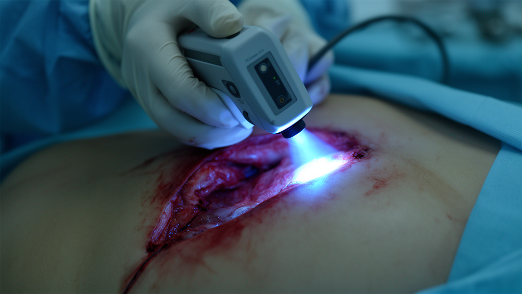
A clinical evaluation of a handheld optical imaging device designated Eagle has demonstrated the intraoperative identification of a grossly occult positive cancer margin during breast conserving surgery. Visualizing malignant tissue in real time remains a persistent clinical obstacle, where positive margins historically drive reoperation rates between 20 and 70 percent under standard white-light illumination and palpation. Conducted across Princess Margaret Cancer Centre and Mount Sinai Hospital in Toronto, the trial evaluated whether the wireless fluorescence device, paired with oral 5-aminolevulinic acid, could reliably discriminate between normal breast architecture and residual carcinoma directly within surgical cavities and on excised specimens. The study aimed to assess clinical utility within active operating room workflows, evaluate ergonomics, and establish the technical baseline required for intraoperative margin assessment.
The biochemical contrast mechanism relies on the selective metabolic accumulation of protoporphyrin IX within cancerous breast tissue following systemic delivery of 5-aminolevulinic acid hydrochloride. As an endogenous non-protein amino acid that participates in mammalian heme biosynthesis, administered 5-aminolevulinic acid is metabolized into heme by cellular pathways throughout the body. However, enzymatic defects inside malignant cells cause abnormal accumulation of the intermediate fluorophore protoporphyrin IX. When illuminated with violet-blue excitation light at wavelengths between 400 and 410 nanometers, accumulated protoporphyrin IX emits red fluorescence with a primary emission peak centered at 635 nanometers. While oral 5-aminolevulinic acid hydrochloride is already approved in approximately forty countries for fluorescence-guided resection of malignant gliomas, this trial investigated its translation to breast conserving surgery using an oral dosage of 20 milligrams per kilogram of body weight taken two to four hours before surgery.
The Eagle system directly addressed several performance bottlenecks identified in earlier clinical research conducted with a predecessor prototype designated PRODIGI. In a previous forty-five-patient randomized controlled trial at Princess Margaret Cancer Centre, the PRODIGI platform verified that 5-aminolevulinic acid-induced fluorescence could detect invasive ductal carcinoma and ductal carcinoma in situ within excised lumpectomies. However, PRODIGI featured a bulky form factor measuring 30 by 14 by 10 centimeters with a mass of 584 grams, preventing clinicians from inserting the device into the confined space of surgical cavities. Furthermore, that earlier device was assembled from consumer-grade camera components, suffered from illumination non-uniformity within enclosed voids, and experienced frequent motion blur caused by one-to-five-second exposure times. Its loose plastic drape also wrinkled across the lens, introducing optical distortions that impaired image interpretation.
To overcome these physical constraints, researchers collaborated with MolecuLight Incorporated to design the Eagle prototype as a wireless, battery-powered imaging instrument engineered specifically for intra-cavity navigation. The Eagle platform has overall bounding dimensions of 17 by 13 by 12 centimeters with a total mass of 557 grams, terminating in a rigid black anodized aluminum shaft measuring 24 millimeters in diameter and 91 millimeters in length. The housing incorporates an internal rechargeable lithium-ion battery, control electronics, and an organic light-emitting diode color touchscreen that provides high contrast and deep black levels under darkened operating room conditions. To simplify optical evaluation, the team fabricated two physically identical units: a fluorescence imaging configuration containing excitation light-emitting diodes and optical emission filters, and a separate white-light configuration featuring dual 5000 Kelvin light-emitting diodes without an emission filter.
The distal imaging tip of the fluorescence configuration integrates optical components tailored specifically to protoporphyrin IX excitation and detection. Excitation illumination is supplied by two 405-nanometer light-emitting diodes with a bandwidth of plus or minus 15 nanometers at full width at half maximum, positioned adjacent to an 8-megapixel complementary metal oxide semiconductor image sensor. Light returning to the camera sensor traverses a custom optical emission filter featuring an optical density of at least 6 between 300 and 492 nanometers to block excitation light. The filter incorporates two distinct transmission bands: a green passband spanning 500 to 545 nanometers and a red passband spanning 600 to 660 nanometers. This dual-band arrangement permits the sensor to capture red fluorescence emitted by protoporphyrin IX in tumors alongside green autofluorescence from collagen in healthy connective tissue, resolving features down to 89 micrometers at a 3-centimeter working distance.
The clinical trial operated under institutional review approvals from the research ethics boards of Princess Margaret Cancer Centre and Mount Sinai Hospital under ClinicalTrials.gov identifier NCT01837225. A total of 32 patients consented to participate, of whom eleven were withdrawn before data collection due to treatment modifications or administrative delays, and four additional patients were withdrawn due to incomplete data acquisition, leaving 17 evaluable patients. Participants received oral 5-aminolevulinic acid hydrochloride at 20 milligrams per kilogram of body weight between two and four hours prior to surgery. Following primary lumpectomy excision, researchers acquired white-light and fluorescence photographs and videos of cavity walls and excised specimens. Targeted cavity and specimen biopsies were cataloged, fixed in formalin, and submitted for comprehensive hematoxylin and eosin histopathologic analysis by an independent study pathologist blinded to imaging results.
Geometric characterization across the 17 surgical cavities confirmed the operational feasibility of navigating the 24-millimeter-wide distal shaft within confined resections. Measured cavities exhibited a mean skin incision length of 5.63 centimeters, an oval opening averaging 4.83 centimeters along the long axis and 4.48 centimeters along the short axis, and an average cavity depth of 3.33 centimeters. Normal breast tissues were differentiated by their fluorescence appearance and quantified using the International Commission on Illumination Delta E 2000 color-difference metric. Fibrous connective tissue appeared bright green under violet illumination, whereas adipose tissue appeared orange-brown. Histopathologic verification confirmed that seven of eight biopsies suspected to be connective tissue contained at least 50 percent connective tissue, with one false reading caused by lymphatic mapping dye Patent Blue V. Similarly, all fourteen biopsies classified as adipose tissue based on fluorescence appearance contained at least 50 percent adipose tissue on microscopic section.
Quantitative colorimetric evaluation demonstrated pronounced visual contrast between malignant tissue and adjacent normal parenchyma across sectioned lumpectomy specimens. Among four evaluated lumpectomy specimens containing invasive ductal carcinoma and ductal carcinoma in situ, the mean Delta E color difference between red fluorescent tumor foci and green connective tissue reached 55.9 plus or minus 14.8, while the color difference between tumor foci and orange-brown adipose tissue measured 17.1 plus or minus 4.4. Most notably, intraoperative fluorescence imaging of an intact lumpectomy specimen identified an unsuspected focal area of bright red fluorescence along the anterior surgical margin that had gone undetected under conventional white-light evaluation. Subsequent histopathologic evaluation confirmed that this fluorescent focus corresponded to ductal carcinoma in situ situated less than 1 millimeter beneath the surgical surface, satisfying the Society of Surgical Oncology and American Society for Radiation Oncology definition of a positive margin.
Clinical deployment also revealed significant optical artifacts originating from the custom sterile drape assembly. The drape utilized an optically transparent polycarbonate window measuring 1.8 millimeters in thickness, exhibiting 92 percent light transmission at normal incidence. However, intrinsic fluorescence from the window material and bonding epoxy introduced a generalized green background tint into captured imagery. Although this green tint mathematically increased quantum dot phantom contrast against pork tissue, fingerprint grease and surface smudges worsened optical distortion. More critically, warm, humid conditions within surgical cavities induced rapid moisture condensation on the cold polycarbonate window within three to five seconds of cavity entry. This fogging caused substantial optical haze, shifting the average cavity color by a Delta E exceeding 40 and reducing the color contrast between adipose and connective tissue by 40.7 to 84.5 percent, although removing the device for two to five seconds allowed the haze to clear.
Illumination characterization exposed substantial non-uniformity across the imaging field of view stemming from the unilateral arrangement of the excitation light-emitting diodes below the camera sensor. Measured optical power density dropped from 71.1 milliwatts per square centimeter at a 3-centimeter working distance to 9.5 milliwatts per square centimeter at 10 centimeters, with illumination heavily concentrated along the bottom edge while upper corners received 3 percent or less of maximum intensity. To eliminate severe vignetting and excitation bleed, engineers cropped the sensor field of view to 68 percent of its linear dimensions, leaving only 46 percent of the usable area. This restriction required imaging excised lumpectomies from 10 centimeters away, reducing optical excitation and rendering faint tumor fluorescence difficult to resolve. Additionally, pooled blood within the cavity strongly absorbed violet excitation light, while specular reflections from red LED spectral leakage mimicked protoporphyrin IX fluorescence.
Operating room observations underscored ergonomic and procedural challenges that extended cavity surveys to between 2 and 2.5 minutes, exceeding the target duration of one minute. Two-handed handling required a second clinician to retract cavity walls, especially when viewing lateral margins where the screen faced away from the operator. Draping demanded a three-person workflow and diminished touchscreen sensitivity, while one-to-two-second exposures risked motion blur and protoporphyrin IX photobleaching. To mitigate these artifacts, researchers proposed preheating the LEDs for two minutes to eliminate condensation, positioning excitation LEDs symmetrically around the sensor to restore field-of-view uniformity, and adding short-pass excitation filters to suppress blood reflections. Incorporating wireless video streaming, physical buttons, and combined white-light and fluorescence imaging will further optimize clinical workflow. Intellectual property licensee SBI ALApharma Canada has begun developing a second-generation Eagle platform incorporating these mitigations to advance intraoperative fluorescence margin detection in future trials.
Subject of Research: Intraoperative fluorescence imaging for breast conserving surgery margin assessment
Article Title: A handheld device for intra-cavity and ex vivo fluorescence imaging of breast conserving surgery margins with 5-aminolevulinic acid
Article References: Gibson, C., Wang, S. C., Phoon, A., Thalanki Anantha, N., Ottolino-Perry, K., Petropoulos, S., Qureshi, Z., Subramanian, V., Shahid, A., O’Brien, C., Carcone, S., Chung, S., Tsui, T., Son, V., Sukhram, M., Meng, F., Done, S. J., Easson, A. M., Cil, T., … DaCosta, R. S. (2024). A handheld device for intra-cavity and ex vivo fluorescence imaging of breast conserving surgery margins with 5-aminolevulinic acid. BMC Biomedical Engineering, 6(1), Article 5. https://doi.org/10.1186/s42490-024-00079-9
Image Credits: AI Generated
DOI: 10.1186/s42490-024-00079-9
Keywords: breast conserving surgery, fluorescence imaging, 5-aminolevulinic acid, protoporphyrin IX, surgical margins, intraoperative imaging, optical diagnostics, lumpectomy, handheld, device, intra-cavity, vivo
News Source: Ophelia Keating. (November 10, 2026). Handheld Fluorescence Device Detects Occult Margins During Breast Conserving Surgery. Scienmag.