Parkinson’s disease may begin influencing the brain long before tremors, stiffness, or slowed movement become visible—and a new study points to an unexpected suspect: inflammatory signals produced by the gut. Researchers Zhang, Zhong, Gao and colleagues report that tumor necrosis factor alpha, or TNF-α, derived from the gut microbiota can activate a molecular pathway that drives the death of dopamine-producing neurons. Their findings, published in Cell Death Discovery, connect the intestinal ecosystem to ferroptosis, a recently recognized form of cell death increasingly associated with neurodegenerative disease.
The study centers on the biological communication system linking the gut and the brain. The gut microbiota consists of trillions of microorganisms that produce metabolites and influence immune activity throughout the body. When this microbial community becomes imbalanced, a condition often called dysbiosis, it can promote chronic inflammation. TNF-α is one of the immune system’s most powerful inflammatory messengers. Although it is essential for fighting infection and coordinating immune responses, excessive or persistent TNF-α signaling can damage tissues, including the nervous system.
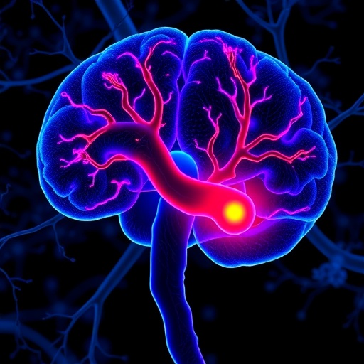
In Parkinson’s disease, the most vulnerable cells are dopaminergic neurons in a midbrain region called the substantia nigra. These neurons release dopamine, a chemical messenger required for smooth, coordinated movement. As they disappear, dopamine levels fall and the characteristic motor symptoms of Parkinson’s emerge. The new research proposes that gut microbiota-derived TNF-α may intensify this process by engaging TNFR1, a cell-surface receptor that detects TNF-α and transmits inflammatory signals into the cell.
According to the researchers, TNFR1 activation initiates a cascade involving NF-κB and ATF4. NF-κB is a transcription factor that controls the expression of numerous genes involved in inflammation, immunity, and cellular stress. ATF4 is another stress-responsive transcription factor, activated when cells struggle with insufficient nutrients, protein-folding problems, oxidative damage, or other forms of metabolic pressure. Together, the TNFR1-NF-κB-ATF4 axis appears to push dopaminergic neurons toward a lethal state rather than allowing them to recover from injury.
That lethal state is ferroptosis. Unlike apoptosis, the orderly form of programmed cell death, ferroptosis is driven by iron-dependent oxidative damage to cell membranes. When reactive oxygen molecules attack polyunsaturated fatty acids in membranes, they initiate a chain reaction known as lipid peroxidation. Normally, antioxidant systems—especially the glutathione and glutathione peroxidase 4 network—keep this chemistry under control. During ferroptosis, those defenses become inadequate, iron helps accelerate the damage, and the membrane eventually loses its integrity.
Dopaminergic neurons may be particularly vulnerable because of their high metabolic demands, extensive branching, and the chemical properties of dopamine itself. Dopamine metabolism can generate reactive molecules, while the substantia nigra naturally contains abundant iron. These factors can create a precarious balance between normal neuronal function and oxidative stress. The study’s proposed mechanism suggests that inflammatory signaling from the gut further weakens this balance, activating cellular stress programs through NF-κB and ATF4 and making ferroptotic damage more likely.
The findings are significant because they unite several major themes in Parkinson’s research: intestinal dysbiosis, systemic inflammation, immune signaling, oxidative stress, and neuronal iron toxicity. Rather than treating these processes as separate contributors, the TNFR1-NF-κB-ATF4 model presents them as connected stages in a biological chain. Gut-derived TNF-α may act as an initiating signal, TNFR1 as the receptor that receives it, NF-κB as an inflammatory amplifier, and ATF4 as a stress-response regulator that helps determine whether a neuron survives or enters ferroptosis.
This mechanism could open new therapeutic possibilities, although it does not yet represent a ready-made treatment. Potential strategies might include reducing harmful inflammatory signaling, selectively blocking TNFR1, modulating NF-κB or ATF4 activity, restoring antioxidant capacity, or protecting neurons from iron-driven lipid peroxidation. Manipulating the gut microbiota is another possibility, but the microbiome is a complex ecosystem and broad interventions can produce unpredictable effects. Any future treatment would need to suppress damaging inflammation without disabling the immune functions required for protection.
The research also highlights why Parkinson’s disease is increasingly viewed as a disorder involving the whole body rather than only the brain. The gut-brain connection may help explain why gastrointestinal symptoms can appear years before classical motor signs in some patients. At the same time, the proposed pathway will require further validation to determine how strongly it operates in human disease, which microbial communities produce the relevant inflammatory signals, and whether interrupting the pathway can preserve dopamine neurons. By identifying a possible molecular bridge from gut-derived TNF-α to ferroptotic neuronal death, the study offers a compelling new framework for understanding—and potentially slowing—the progression of Parkinson’s disease.
Subject of Research: Gut microbiota-derived TNF-α, ferroptosis, and dopaminergic neuron loss in Parkinson’s disease
Article Title: Gut microbiota-derived TNF-α triggers dopaminergic neuron ferroptosis via TNFR1-NF-κB-ATF4 axis in Parkinson’s disease
Article References: Zhang, Z., Zhong, S., Gao, L. et al. “Gut microbiota-derived TNF-α triggers dopaminergic neuron ferroptosis via TNFR1-NF-κB-ATF4 axis in Parkinson’s disease.” Cell Death Discovery (2026). https://doi.org/10.1038/s41420-026-03281-x
Image Credits: AI Generated
DOI: https://doi.org/10.1038/s41420-026-03281-x
Keywords: Parkinson’s disease, gut microbiota, TNF-α, ferroptosis, dopaminergic neurons, TNFR1, NF-κB, ATF4, neuroinflammation, iron-dependent cell death
Tags: dysbiosis and neuroinflammatory pathwaysearly biomarkers of Parkinson’s diseaseferroptosis in dopaminergic neuronsgut microbiota and neuroinflammationgut microbiota influence on neurodegenerative diseasesgut-brain axis and neurodegenerationimmune mechanisms underlying neuron ferroptosismicrobial metabolites and immune responserole of inflammatory cytokines in Parkinson’sTNF-α in Parkinson’s diseaseTNFR1-NF-κB-ATF4 signaling in neuron death