
Investigating the remarkable structure and function of the zebrafish retina has recently shed light on how photoreceptors, the essential cells responsible for vision, maintain their precise arrangement within this critical organ. Researchers at the esteemed Okinawa Institute of Science and Technology (OIST) have unveiled the role of a protein known as Dscamb in organizing cone cells in the retina, which is significant for optimal visual acuity. The cone mosaic, characterized by an orderly layout of different cone types responsible for color vision, relies heavily on this regulatory mechanism. This breakthrough presents new avenues for understanding the neural mechanisms underlying visual processing and spatial organization in vertebrate retinas.
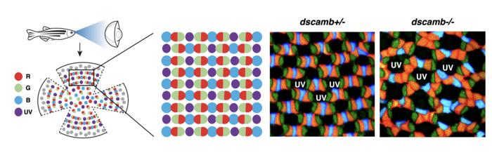
The retinal structure of zebrafish is particularly fascinating due to its highly specialized photoreceptor cells. Cones are essential for daylight vision and color detection, and zebrafish possess an impressive variety comprising red, green, blue, and UV-sensitive cones. This diversity allows for exceptional color discrimination in a species known for its agility and acuity within aquatic environments. The patterned arrangement of these cones within a mosaic is not arbitrary; rather, it is a function of complex cellular interactions that ensure optimal spacing and functionality. The discovery of Dscamb’s role could be pivotal in unraveling the molecular foundation of these spatial arrangements.
The term “cone mosaic” refers to the intricate ordering of cone cells within the retina, a phenomenon characterized by remarkable regularity. Unlike a random distribution, these cells are arranged such that cones of the same type maintain specific distances from one another and create discernible patterns with adjacent types. The evolution of this precise spatial organization has remained a mystery in vision science for decades. The recent findings by the OIST team provide a critical piece of the puzzle, articulating a molecular mechanism that governs these arrangements across various vertebrate species.
Dscamb, or Down Syndrome Cell Adhesion Molecule-B, is part of a larger family of proteins known to facilitate cell-cell adhesion during the development of the nervous system. Each vertebrate species has evolved distinct versions of this protein, which serve similar functions in diverse contexts. In zebrafish, Dscamb plays a unique role, located specifically within the developing photoreceptors of the retina, pointing to its specialized function in ensuring the correct formation of the cone mosaic.
To ascertain the function of Dscamb in cone cell arrangement, researchers conducted genetic modifications in zebrafish to create mutants that lack this essential protein. Upon examination, the resulting cone mosaics revealed striking abnormalities, particularly noticeable clustering of red cone cells. The findings demonstrated that Dscamb is indispensable in maintaining the symmetric and balanced distribution of cones, particularly in red cones, emphasizing its role as a critical self-avoidance enforcer.
Through rigorous experimentation and analysis, it became clear that the behavior of cone filopodia—a mechanism of cellular protrusions—plays an important part in this process. The zebrafish cone photoreceptors extend these projections to establish contact with neighboring cones during early developmental stages. Interestingly, filopodia only retract when contacting the same type of cone, indicating a recognition system at work that relies on Dscamb. This suggests an intrinsic ability for cone cells to “sense” the presence of like cells and respond appropriately, thus reinforcing the spatial integrity of the mosaic.
Further investigations revealed that this self-avoidance mechanism is fine-tuned to subtypes of cone cells. For instance, red cones demonstrate a distinctive repulsive response towards one another via Dscamb interactions, while the spacing between blue cones appears to be regulated by different mechanisms. Such specificity in cone-type recognition may provide essential insights into how the visual system maintains clarity and precision, despite the diverse array of cone types present.
The implications of these discoveries extend beyond zebrafish and into broader fields of vision research. Understanding the molecular orchestrations that underlie visual mechanisms is pivotal for uncovering the complexities of retinal disorders in humans. The parallels drawn from photoreceptor spacing can lead to potential advancements in treatment methodologies for retinal diseases often associated with disrupted cellular organization, such as age-related macular degeneration and retinitis pigmentosa.
Moreover, this research opens up exciting avenues for innovation in regenerative medicine. Insights gained from the zebrafish model, particularly regarding photoreceptor development and spatial regulation, could inform therapeutic strategies aimed at restoring vision in individuals suffering from retinal degenerative conditions. The knowledge of how cells communicate and form organized structures can be harnessed for producing bioengineered tissues that replicate natural ocular responses.
As a testament to the vibrant scientific discourse around this groundbreaking work, the research findings have been published in the distinguished journal Nature Communications. The study not only highlights the role of Dscamb in photoreceptor organization but also sets the stage for further inquiry into molecular patterns governing cell arrangements across various biological systems. These findings exemplify an essential step forward, bridging knowledge gaps in our understanding of vision science while illuminating potential pathways for clinical application.
The study, led by Dr. Dongpeng Hu, a former PhD student at OIST’s Developmental Neurobiology Unit, emphasizes the crucial interplay between molecular mechanisms and the formation of complex biological structures. Dr. Hu’s dedication to elucidating these fundamental processes is reflective of a broader mission within the scientific community—to decode the intricate language of cell interactions and their implications for health and disease.
Undoubtedly, the revelations surrounding Dscamb and its role in cone mosaic development represent a foundational shift in biomedical research. This newfound understanding illuminates the underlying principles of cellular assembly and positioning, transforming not only the landscape of vision science but also informing the discourse on developmental biology as a whole. Researchers are now equipped with a conceptual toolkit for exploring analogous mechanisms across various tissues, fostering a richer grasp of life’s complexity at a cellular level.
The combined expertise of the OIST team and their innovative approach to investigating these phenomena exemplifies the spirit of scientific inquiry. They have not only contributed to a nuanced understanding of retinal morphology but have also catalyzed a discussion about the future of vision research—a discussion that holds tremendous promise for enhancing our comprehension of both normal and pathologic visual processes.
By unraveling the intricacies behind how retinal cells maintain their distance and organize into functional patterns, researchers venture further into understanding the delicate balance that governs the development and function of complex tissues. This study serves as a clarion call to scientists, signaling the importance of molecular research in revealing the secrets of form and function in the living world, ultimately driving progress towards therapies that can transform lives.
Subject of Research: Animals
Article Title: Study shows how retinal cells know when to keep their distance
News Publication Date: 25-Mar-2025
Web References: https://www.nature.com/articles/s41467-025-57506-1
References: DOI 10.1038/s41467-025-57506-1
Image Credits: Credit: Hu et al., 2025
Keywords
Tags: advancements in retinal researchcellular interactions in retinal mosaicscone cell organization in retinaimplications for understanding vision disordersmechanisms of retinal cell spacingneural mechanisms of visionoptimal visual acuity and color visionphotoreceptor diversity in zebrafishrole of Dscamb protein in visionsignificance of cone mosaic arrangementvisual processing in vertebrateszebrafish retinal structure

