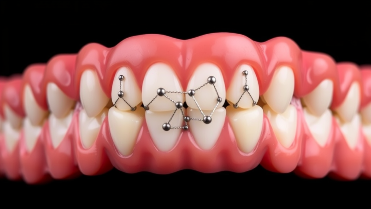
Dental implants have become one of the most predictable long-term solutions for tooth replacement, yet their success hinges on a deceptively simple biological event: the direct, functional connection between the implant surface and the surrounding jawbone, known as osseointegration. Titanium, the workhorse material of implant dentistry, offers excellent mechanical strength and corrosion resistance, but its naturally bioinert surface limits direct biological interaction with bone tissue. When early bone-implant integration falters, implants can loosen and fail. To overcome this, researchers have long turned to hydroxyapatite (HA), a calcium phosphate mineral chemically akin to natural bone, as a bioactive coating that encourages bone-forming cells to attach and deposit new matrix. Clinical experience, however, has revealed persistent weaknesses: plasma-sprayed HA coatings can delaminate over time, and poorly crystalline layers may resorb rapidly while binding proteins inefficiently.
Now, a study published in the Journal of Cellular and Molecular Medicine proposes a molecularly grounded strategy for upgrading these coatings. A research team from King Khalid University and collaborating institutions investigated what happens at the atomic scale when graphene oxide (GO), a chemically functionalised derivative of graphene, is combined with hydroxyapatite in a nanocomposite surface. Using an integrated suite of computational techniques—molecular docking, molecular dynamics simulations, three-dimensional pharmacophore mapping, and in silico toxicity screening—the researchers systematically compared how extracellular matrix (ECM) proteins and integrin receptors engage with pure HA, pure GO, and the combined GO–HA surface. Their central question: does graphene oxide genuinely enhance the molecular dialogue that sparks bone formation at the implant interface?
The choice of biological players was deliberate and biologically grounded. The team assembled a panel of twelve ECM proteins central to bone regeneration, including fibronectin, collagen type I, laminin, periostin, vitronectin, osteocalcin, osteonectin, bone sialoprotein, osteopontin, biglycan, decorin, and tenascin-C. Each plays a defined role in osseointegration—fibronectin drives osteoblast adhesion and migration through its RGD peptide motifs, collagen type I forms the primary organic scaffold of bone, and proteoglycans such as decorin and biglycan orchestrate collagen fibril assembly and growth factor activity. On the receptor side, six integrins were modelled, including α5β1, the principal fibronectin receptor, αvβ3, which binds RGD-rich bone proteins, and α2β1, the major collagen receptor. These transmembrane molecules are the mechanistic gateway through which cells sense a biomaterial surface and launch the signalling cascades leading to survival, proliferation, and osteogenic differentiation.
The docking results delivered a clear message. While hydroxyapatite showed respectable binding to laminin—its best-performing complex, with a HADDOCK score of −19.7 and a binding free energy of −4.95 kcal/mol—the graphene oxide-containing systems outperformed HA alone by a wide margin. The GO–α5β1 integrin complex posted a HADDOCK score of −56.0 and a striking binding affinity of −10.53 kcal/mol, driven by powerful van der Waals (−33.7 kcal/mol) and electrostatic (−53.3 kcal/mol) contributions and the largest buried surface area in the study at 745.4 square angstroms. GO complexes with α6β1 and laminin were similarly strong. The investigators attribute this enhanced interaction potential to GO’s exceptional mechanical strength, vast surface area, and dense array of oxygen-containing functional groups—hydroxyl, carboxyl, and epoxide moieties—that fuel hydrogen bonding and polar contacts with target proteins.
The most impressive performance came from the hybrid surface. The GO_HA_α5β1 complex achieved the strongest docking score of the entire study, −71.4 ± 2.2, with a binding free energy of −12.33 kcal/mol and the largest buried surface area recorded, 915.3 square angstroms. Specific atomic contacts helped explain the synergy: a hydrogen bond formed between a GO hydroxyl group and the Tyr287 residue of the integrin, reinforced by attractive charge interactions with Arg350, van der Waals forces, and π–π stacking against the aromatic carbon backbone. Complexes with periostin and vitronectin reached ΔG values of −11.63 and −10.93 kcal/mol, respectively, both exceeding anything achieved with pure hydroxyapatite. Intermolecular contact analysis reinforced the picture, with the GO_HA_α5β1 system registering thousands of carbon-carbon and carbon-oxygen atom-pair contacts—markers of extensive hydrophobic and polar interaction networks across a broad molecular interface.
Pharmacophore mapping added chemical nuance to the binding story. For the α5β1 complex, clusters of hydrogen-bond acceptors gathered around GO’s epoxide and hydroxyl groups, while a dense web of hydrogen-bond donors—traced largely to HA’s phosphate and calcium-bound hydroxyl sites—lined the opposite side of the interface. The periostin complex displayed a balanced, evenly distributed network of donors and acceptors that the authors describe as an effective molecular anchor, while vitronectin’s β-sheet regions aligned with hydrophobic patches along the GO aromatic framework. In essence, the two materials divide the labour: graphene oxide supplies electron-rich acceptor chemistry and hydrophobic surface anchoring, while hydroxyapatite contributes polar donor sites, producing a reactive, dual-function surface that ECM proteins grip more firmly than either component alone.
Molecular dynamics simulations running 100 nanoseconds under physiological conditions—310 K, 1 bar, explicitly solvated—tested whether these docked encounters survive thermal motion. Across three flagship complexes (α5β1 integrin, periostin, and vitronectin), the GO–HA assemblies consistently showed the highest numbers of hydrogen bonds and the lowest residue-level fluctuations. The GO_HA_periostin complex, for instance, formed 20 hydrogen bonds—double the count of the HA-only equivalent—and recorded the lowest root-mean-square fluctuation among all periostin systems at 0.852 nm, indicating that individual residues were held more tightly in place. Similarly, GO_HA_vitronectin stabilised the functionally important Ser180–Phe210 region and maintained 19 hydrogen bonds. While the hybrid complexes exhibited larger radii of gyration, reflecting an extended conformation that embraces more surface contact, the combined evidence points to a surface that holds proteins in stable, potentially more bioactive orientations rather than merely sticking them down loosely.
Safety, inevitably, complicates the enthusiasm. In silico toxicity screening painted a stark contrast between the two components: hydroxyapatite showed no predicted mutagenicity, tumorigenicity, irritancy, or reproductive toxicity, consistent with its long clinical record in bone grafting, whereas graphene oxide triggered high-risk flags across all four endpoints. GO’s intermediate lipophilicity, large hydrophobic surface area, and predicted capacity to cross cellular barriers underlie these warnings. Experimental literature offers both caution and a remedy: smaller GO nanosheets can be internalised by cells, provoking oxidative stress and membrane damage, yet embedding GO within a calcium phosphate matrix may neutralise its excessive negative surface charge, mask reactive oxygen functionalities, and temper reactive oxygen species generation. The authors are explicit that this protective effect remains a theoretical postulate requiring cytotoxicity assays and in vivo validation before any clinical claim can be made.
The team is equally candid about the boundaries of computational prediction. Their model represents GO and HA as component-level molecular structures within a simplified interfacial assembly—it does not validate a covalent GO–HA bond, coating morphology, or crystallinity, and it cannot capture competitive serum protein adsorption, immune responses, biomechanical loading, or the messy temporal dynamics of living bone. The findings are framed as a framework for generating testable hypotheses rather than proof of clinical efficacy. The validation roadmap is nonetheless concrete: surface characterisation by atomic force microscopy, electron microscopy, and X-ray photoelectron spectroscopy; protein adsorption assays; osteoblast cell-culture experiments measuring adhesion, viability, and osteogenic differentiation; macrophage and cytokine profiling to rule out adverse immunological reactions; and eventually animal implantation studies assessed by micro-computed tomography, histomorphometry, and pull-out testing.
If those experiments confirm what the simulations suggest, the implications extend well beyond dentistry. A coating that simultaneously strengthens the mechanical integrity of the implant interface, amplifies protein adsorption, exposes integrin-binding domains in adsorbed fibronectin, and passes biological safety screening would address the central weakness of titanium implants: their silence in the molecular language of bone. The study’s real contribution is a rigorous, multi-method template for evaluating next-generation biomaterial surfaces before a single laboratory experiment is run—accelerating the search for implant coatings that coax bone to embrace metal not reluctantly, but eagerly.
Subject of Research: Computational analysis of graphene oxide–hydroxyapatite nanocomposite coatings and their interactions with extracellular matrix proteins to improve dental implant osseointegration.
Article Title: Graphene Oxide–Hydroxyapatite Nanocomposite Coatings and Extracellular Matrix Protein Interactions for Enhanced Osseointegration in Dental Implants
Article References: Saini, R. S., Binduhayyim, R. I. H., Dermawan, D., Kanji, M. A., Quadri, S. A., & Heboyan, A. (2026). Graphene Oxide–Hydroxyapatite Nanocomposite Coatings and Extracellular Matrix Protein Interactions for Enhanced Osseointegration in Dental Implants. Journal of Cellular and Molecular Medicine, 30(17), Article e71352. https://doi.org/10.1111/jcmm.71352
Image Credits: AI Generated
DOI: 10.1111/jcmm.71352
Keywords: dental implants, osseointegration, graphene oxide, hydroxyapatite, nanocomposite coatings, extracellular matrix proteins, molecular docking, molecular dynamics simulation, integrin receptors, biomaterials, in silico toxicity, titanium implants
Cite Scienmag News
APA MLA Chicago
Neil Sanderson. (September 13, 2026). Graphene-Infused Coatings May Help Dental Implants Bond Better to Bone. Scienmag. https://scienmag.com/graphene-infused-coatings-may-help-dental-implants-bond-better-to-bone/
Neil Sanderson. “Graphene-Infused Coatings May Help Dental Implants Bond Better to Bone.” Scienmag, 13 September 2026, https://scienmag.com/graphene-infused-coatings-may-help-dental-implants-bond-better-to-bone/. Accessed 13 September 2026.
Neil Sanderson. “Graphene-Infused Coatings May Help Dental Implants Bond Better to Bone.” Scienmag. September 13, 2026. https://scienmag.com/graphene-infused-coatings-may-help-dental-implants-bond-better-to-bone/
Copy citation Download RIS
Tags: advanced coatings for dental implantsbioactive implant coatingsbioinert titanium surface modificationbiomaterialsbone-implant bonding improvementDental implant surface modificationdental implantsextracellular matrix proteinsgraphene oxidegraphene oxide hydroxyapatite nanocompositegraphene-based biomaterialsgraphene-infused dental materialshydroxyapatiteimplant failure prevention strategiesin silico toxicityintegrin receptorsmolecular dockingmolecular dynamics simulationmolecular modeling of implant surfacesnanocomposite coatingsnanotechnology in dentistryosseointegrationosseointegration enhancementtitanium implants