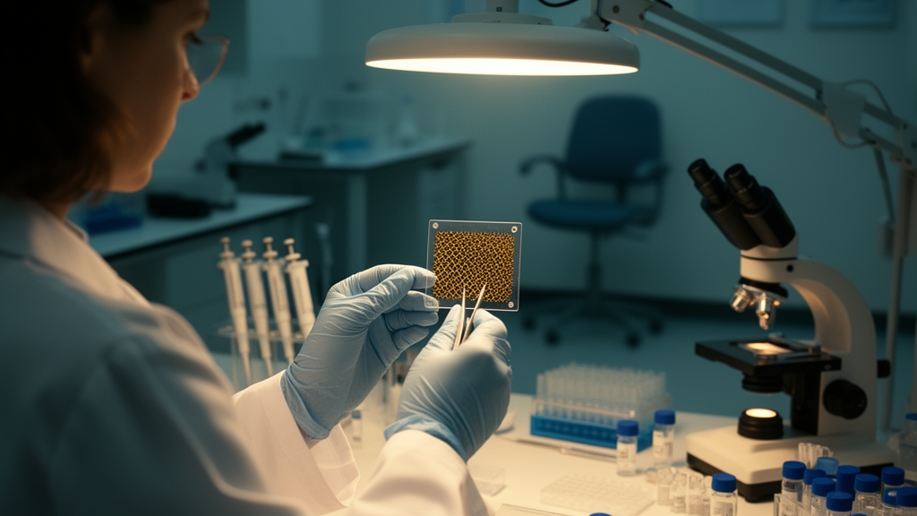
Graphene has spent two decades being billed as the wonder material that will shrink laboratory diagnostics onto a chip, a strip of paper, or even a contact lens. A new open-access review in Discover Nano takes that promise seriously enough to interrogate it, line by line, and its verdict is more sobering than the field’s usual hype. The authors, led by Hamza Rafeeq and Huanhuan Chen, survey graphene oxide, reduced graphene oxide, graphene quantum dots, and graphene-based composites across optical, electrochemical, and transistor-based sensing platforms, and conclude that while the analytical chemistry is often spectacular, the path to the clinic is blocked less by sensitivity than by reproducibility, standardisation, and validation.
The review’s central critical finding concerns the headline numbers that dominate the graphene biosensing literature: femtomolar and attomolar detection limits. The authors point out that most such figures are calculated as three times the standard deviation of the blank divided by a calibration slope, using curves whose lowest measured concentration sits two to four orders of magnitude above the quoted limit. In other words, the detection limit is an extrapolation, not an observation. The authors also flag a physical problem: at one attomolar in a ten-microlitre droplet, the assay volume contains roughly six target molecules, and diffusion-limited capture of even a meaningful fraction of those would take hours, not the minutes such assays report. Very few papers state the assay volume alongside the limit at all.
Validation is the third recurring weakness. Real-sample performance is typically assessed by spike-and-recovery experiments, in which purified analyte is added to pooled serum, urine, milk, or juice and recoveries between 90 and 110 percent are reported. That design tests for gross matrix interference and nothing more: it does not test endogenous analyte in its native form, does not sample biological variability across individuals, and is not blinded. Genuine clinical validation would require blinded patient specimens analysed against a reference method, with agreement assessed by Bland–Altman or Passing–Bablok regression and discrimination reported as sensitivity, specificity, and area under the receiver-operating-characteristic curve. The authors find compliance with the STARD reporting guideline for diagnostic accuracy studies close to absent in this literature.
The material itself is part of the problem. Graphene oxide is not a stoichiometric compound but an amorphous, non-periodic solid whose carbon-to-oxygen ratio varies with synthesis conditions, so “GO” describes a family of materials rather than a single substance. A systematic survey of commercially supplied graphene powders from producers across three continents, cited in the review, found that no sample contained more than roughly half graphene by content, that most consisted predominantly of graphite, and that some carried substantial contamination. Certified reference materials for graphene do not yet exist, which means inter-laboratory reproducibility of graphene biosensors is bounded above by the reproducibility of the starting material, which the authors describe as poor.
Even the electrochemistry has a hidden confound. Graphene prepared by oxidative routes retains trace manganese from permanganate oxidants, and chemical-vapour-deposition graphene retains copper or nickel from the growth catalyst. The review highlights work showing that such metallic impurities, present at parts-per-million levels and invisible to routine characterisation, can dominate the apparent electrocatalytic response toward hydrogen peroxide and related analytes, producing signals attributed to the carbon lattice that are in fact metallic in origin. The authors recommend that reports of intrinsic graphene electrocatalysis be supported by impurity quantification, for example by inductively coupled plasma mass spectrometry, and benchmarked against edge-plane and basal-plane pyrolytic graphite controls.
Field-effect transistor biosensors, the most hyped corner of the field, face their own electrostatic ceiling. In physiological media of roughly 150 millimolar ionic strength, the Debye screening length is only about 0.7 to 0.8 nanometres, so charge from a biomolecule bound beyond that distance produces almost no change in channel conductance. Since an immunoglobulin G molecule is roughly 10 to 15 nanometres across, a target captured at the tip of a full-size antibody is largely invisible to the transistor. This, the authors argue, is why many reported graphene-FET assays are run in diluted buffer rather than undiluted serum or saliva. Baseline drift of tens of millivolts per hour, non-specific adsorption of serum proteins onto graphene’s hydrophobic surface, and wafer-scale fabrication spreads exceeding one hundred millivolts in Dirac-point voltage compound the problem, forcing per-device calibration that is incompatible with single-use point-of-care operation.
The review also corrects some widely repeated claims. The seminal graphene-FET report on SARS-CoV-2, while genuinely detecting virus in clinical swab samples, established a limit of detection of 2.42 × 10² copies per millilitre in transport medium; the attomolar figures quoted elsewhere derive from purified spike-protein spiking experiments in dilute buffer, and the two should not be conflated. Similarly, the frequently cited “up to 50 percent” sensitivity gain for graphene-coated SPR chips is a simulation-derived figure for an optimised number of layers; experimentally, the improvement is layer-number dependent and can become negative beyond a few layers as optical absorption broadens the plasmonic resonance. And the claim of direct electron transfer between glucose oxidase and graphene electrodes is, in the authors’ assessment, in most cases attributable to free or adsorbed FAD released from denatured enzyme, a redox-active but catalytically incompetent species.
None of this diminishes the genuine strengths. Enzymatic electrochemical sensing emerges as the most translationally mature modality, already embodied in commercial glucometers and continuous glucose monitors, with screen-printable electrodes and handheld potentiostats. Affinity-based electrochemical sensors, particularly aptasensors on screen-printed graphene, are identified as the most probable near-term route to a clinically deployed graphene diagnostic, combining sub-picomolar sensitivity with the manufacturing base of the glucose-strip industry. Graphene-FET and SERS platforms deliver the most extreme reported sensitivities but remain the least reproducible and least manufacturable, their figures of merit being analytical ceilings under idealised conditions rather than performance that can currently be relied upon in a clinical workflow.
The regulatory picture is stark. In the European Union, the In Vitro Diagnostic Regulation, in force since May 2022, assigns devices to risk classes A to D and requires notified-body involvement and a performance-evaluation report for the great majority of formerly self-certified assays. In the United States, a novel biosensor without a legally marketed predicate cannot use the 510(k) route and must proceed by De Novo classification or premarket approval, while a CLIA waiver for decentralised use requires a separate demonstration that untrained operators obtain equivalent results. Manufacture must occur under an ISO 13485 quality management system mandating a defined, auditable specification for every incoming material, precisely the specification that cannot presently be written for graphene. To the best of the authors’ knowledge, no graphene-based biosensor had obtained FDA clearance or CE-IVD certification under the IVDR at the time of writing, despite two decades of research and several thousand publications.
The authors propose a minimum reporting standard: state the assay volume and incubation time alongside any detection limit, distinguish extrapolated limits from experimentally verified quantification limits, report inter-device and inter-batch relative standard deviations with the number of independent devices, quantify metallic impurities where electrocatalytic enhancement is claimed, evaluate performance in undiluted matrix or state the dilution factor explicitly, and validate against a reference method in blinded patient specimens with STARD-compliant accuracy metrics. Until such practices become normative, they write, the exemplary values quoted throughout the field, including the 0.02 fM miRNA, 1.90 fg/mL CA 15-3, and attomolar SARS-CoV-2 figures, are best interpreted as upper bounds on analytical capability under idealised conditions rather than as demonstrated clinical performance. The binding constraint on progress, they conclude, is no longer sensitivity but reproducibility, standardisation, and validation, and the field should come to value a rigorously validated picomolar assay above an unvalidated attomolar one.
Subject of Research: A critical review of graphene-based nanomaterial biosensors, their reported performance, validation gaps, and barriers to clinical translation.
Article Title: Graphene-based nanomaterials in biosensing: current advances, challenges, and future directions for precision diagnostics
Article References: Rafeeq, H., Khokhar, A. M., Siddique, R., Zia, M. A., Naeem, M., & Chen, H. (2026). Graphene-based nanomaterials in biosensing: current advances, challenges, and future directions for precision diagnostics. Discover Nano, 21(1), Article 552. https://doi.org/10.1186/s11671-026-05057-9
Image Credits: AI Generated
DOI: 10.1186/s11671-026-05057-9
Keywords: graphene, biosensors, graphene oxide, graphene quantum dots, field-effect transistors, SERS, electrochemical sensing, detection limits, clinical validation, point-of-care diagnostics, regulatory translation, nanotechnology
News Source: Neil Sanderson. (October 28, 2026). Graphene biosensors dazzle in the lab but stumble before the clinic. Scienmag.