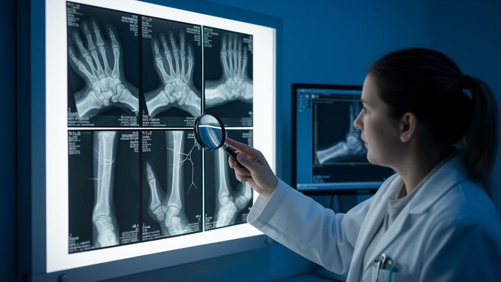
Artificial intelligence systems that read medical images are usually celebrated for what they catch. Far less attention is paid to what they quietly miss. A new study from Ankara Sincan Training and Research Hospital in Türkiye has done exactly that, mapping the failures of a multimodal large language model, the GPT-5 API, when it was asked to detect fractures around the growth plates of children. The results, published in BMC Medical Imaging, are sobering: in 221 of 805 confirmed physeal fractures, the model failed to produce a qualifying fracture call in the correct anatomical region, a false-negative rate of 27.5 percent. For a technology often pitched as a safety net for busy emergency departments, the message is blunt. A negative answer from the model should never be used to rule out a growth plate injury.
The study focused on physeal fractures, injuries that involve the physis, the cartilaginous growth plate found only in children whose skeletons are still developing. These fractures matter enormously. The growth plate is the engine of longitudinal bone growth, and trauma to it can disrupt that process permanently, leading to limb-length discrepancy, angular deformity, or arrested growth if the injury is not recognized and managed correctly. Radiographic assessment is notoriously difficult because cartilage is invisible on plain X-rays, so the fracture line may be subtle, displaced, or entirely occult. Salter-Harris type I injuries, in which the fracture passes through the physis itself, are particularly treacherous, since the only clue may be soft-tissue swelling or a barely perceptible widening of the growth plate.
The research team designed a retrospective, single-center, case-only analysis. They assembled 805 confirmed physeal fractures in children aged zero to twelve years, all sustained during 2025. Each case was evaluated once, in March and April 2026, through a recorded GPT-5 API workflow. The model received anonymized radiographs together with a small, structured package of clinical context: the child’s age, the target anatomical region, and a brief complaint or mechanism of injury. Direct identifiers, accession numbers, exact dates, and burned-in text were removed or masked before submission, in compliance with institutional approval and the Turkish Personal Data Protection Law. The model outputs were generated retrospectively and never influenced the clinical care of any patient.
The reference standard was deliberately conservative. Three orthopedic surgeons, a pediatric orthopedics specialist, an orthopedic trauma specialist, and a senior orthopedic surgeon, each with at least eight years of clinical experience, independently reviewed every radiograph. They then reached consensus with the model outputs concealed, and no radiologist took part. The primary endpoint was a definite or probable fracture call in the correct anatomical region. Importantly, the study did not require the model to explicitly recognize the physis or to assign a correct Salter-Harris classification; simply flagging a fracture in the right place was enough to count as a success. This design choice means the reported sensitivity of 72.5 percent is, if anything, a generous measure of the model’s performance.
Because every case in the cohort was fracture-positive, the study could not estimate specificity, predictive values, overall accuracy, or receiver operating characteristic measures. That is a structural consequence of the case-only design, and the authors are explicit about it. What the design does allow is a clean, unobstructed view of false negatives, the failures that matter most in a screening context. When a model says no and the patient actually has a fracture, the consequences can be serious: delayed presentation, inappropriate discharge, and, in the case of physeal injuries, potential permanent growth disturbance. Understanding where and why those failures cluster is the first step toward mitigating them.
The headline finding was the overall sensitivity of 72.5 percent, with a 95 percent confidence interval of 69.4 to 75.5 percent. But the more interesting story lies in the stratified patterns. In an unadjusted secondary comparison, the false-negative rate was 29.9 percent for Salter-Harris type I and II injuries, compared with 17.9 percent for type III and IV injuries, a risk ratio of 1.67 with a confidence interval of 1.15 to 2.41 and a p-value of 0.004. When type I injuries were excluded entirely, sensitivity barely moved, rising only marginally to 72.8 percent. The pattern is consistent with a known weakness of image-based models: injuries whose radiographic signature is subtle or indirect, such as fractures confined to or passing through the radiolucent growth plate, are harder to detect than injuries with obvious cortical disruption.
Anatomical location also mattered. The descriptive false-negative rates were highest in the elbow, at 36.9 percent, and at the distal tibia, at 31.2 percent. The distal radius, one of the most common sites of pediatric physeal fracture and often the most radiographically obvious, fared better, with a false-negative rate of 20.1 percent. The elbow is a notoriously complex region, with multiple ossification centers that appear and fuse in a predictable sequence as a child grows, and distinguishing a normal ossification center from a fracture fragment requires sophisticated pattern recognition. The distal tibia presents its own challenges, with overlapping bony structures and subtle physeal widening that can evade even experienced eyes.
Age, by contrast, did not significantly change the failure rate. Children aged zero to six years had a false-negative rate of 30.0 percent, while those aged seven to twelve had a rate of 24.9 percent, a difference that was not statistically significant with a p-value of 0.108. This is a hypothesis-generating observation rather than a definitive conclusion, but it suggests that the model’s difficulties are driven more by the nature of the injury and the anatomy involved than by the developmental stage of the skeleton. Younger children do have more cartilage and fewer ossified structures, which might have been expected to make detection harder, yet the difference did not reach statistical significance in this cohort.
The authors are careful to delineate the limits of what their study can claim. It did not assess explicit physeal recognition or Salter-Harris classification, and it cannot establish full diagnostic accuracy, performance relative to clinicians, or clinical utility. There was no comparison group of human readers, so the study cannot say whether the model is better or worse than the clinicians it might assist. The single-center design and the restriction to confirmed fracture cases mean the findings describe failure patterns in a specific population rather than real-world screening performance. Still, the central conclusion stands on its own: under the recorded context-assisted GPT-5 API workflow, more than a quarter of confirmed physeal fractures did not receive a qualifying regional fracture call.
What does this mean for the deployment of multimodal large language models in pediatric imaging? The study is a caution against treating these systems as diagnostic oracles, particularly in the high-stakes domain of growth plate injuries, where a missed fracture can alter a child’s skeletal future. The regional and Salter-Harris stratifications offer actionable intelligence: clinicians and developers now know that type I and II injuries, elbow fractures, and distal tibial fractures are the model’s weakest zones, and that vigilance should be highest exactly where the model is least reliable. As multimodal AI continues its rapid advance into radiology workflows, studies like this one, which map failure patterns with the same rigor usually reserved for successes, will be essential to ensuring that the technology augments human judgment rather than quietly undermining it.
Subject of Research: Artificial intelligence models can miss subtle pediatric growth plate fractures, and a new study maps exactly where the failures cluster.
Article Title: Regional fracture-detection false-negative patterns of a multimodal large language model in confirmed pediatric physeal fractures: a case-only analysis
Article References: Erginoğlu, S. E., Ülgen, N. K., Nazlıgül, A. S., Yiğit, N., & Akkurt, M. O. (2026). Regional fracture-detection false-negative patterns of a multimodal large language model in confirmed pediatric physeal fractures: a case-only analysis. BMC Medical Imaging. https://doi.org/10.1186/s12880-026-02771-9
Image Credits: AI Generated
DOI: 10.1186/s12880-026-02771-9
Keywords: Regional, fracture-detection, false-negative, patterns, multimodal, large, language, model, confirmed, pediatric, physeal, fractures
News Source: Ophelia Keating. (October 19, 2026). GPT-5 Missed More Than a Quarter of Confirmed Pediatric Growth Plate Fractures on X-rays. Scienmag.