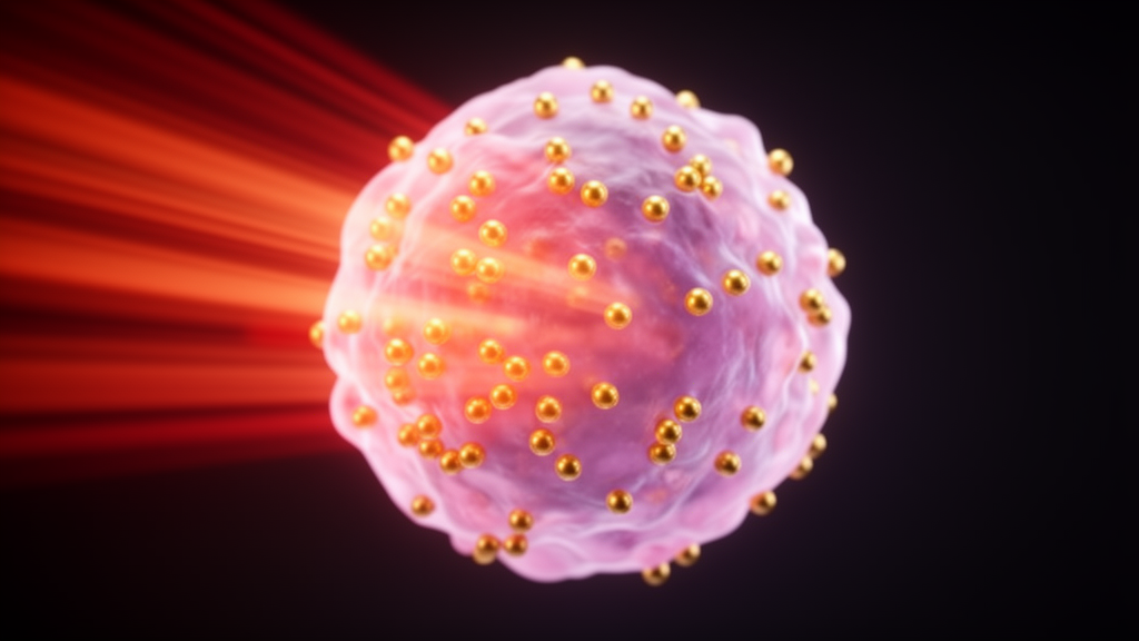
Gold nanoparticles are emerging as one of the most versatile platforms in cancer research, capable of acting simultaneously as imaging contrast agents, drug carriers and heat-generating tumor killers, according to a comprehensive review published in Gold Bulletin. The review, led by Shivanshu Choudhary and Deepak N. Kapoor of Chitkara University with collaborators including Deepak B. Pemmaraju, synthesizes years of laboratory and animal studies to argue that these tiny gold structures could unify cancer diagnosis and treatment in a single theranostic system. The authors emphasize that gold nanoparticles combine unique optical properties, biocompatibility and easy chemical modification, making them unusually adaptable to the diverse demands of oncology.
The clinical backdrop is stark. The review notes that global cancer diagnoses reached more than 19.6 million in 2020, with an estimated 10 million deaths, and projections suggest that by 2040 there will be over 28 million new cases and 16 million fatalities worldwide. Conventional treatments such as chemotherapy often lack selectivity and drug resistance emerges quickly, which drives the search for targeted approaches. Gold has a long medicinal history, having been used by Eastern physicians since antiquity and recognized by Jacques Forestier as an anti-inflammatory for rheumatoid arthritis in the early twentieth century, but it is the nanoscale form that has captured modern attention.
Gold nanoparticles range from roughly 1 to 150 nanometers in core diameter, and their defining optical feature is surface plasmon resonance, a phenomenon in which conduction electrons oscillate in response to light and produce distinct color changes depending on particle size. Smaller particles reflect red light while larger ones absorb it at different wavelengths, a property that underpins both imaging and light-based therapy. Their inherently negative surface charge allows convenient attachment of drugs, targeting ligands, genes and antibodies. Synthesis routes include top-down methods such as laser ablation and ion sputtering, bottom-up reduction of gold ions with sodium citrate or sodium borohydride, the Turkevich method yielding 15 to 30 nanometer spheres, and the Brust-Schiffrin two-phase technique producing particles between 1.5 and 5.2 nanometers. Characterization relies on transmission electron microscopy, dynamic light scattering, zeta potential measurements and UV-Vis spectroscopy.
On the therapeutic side, the review catalogs a striking array of functionalized particles tested in cell and animal models. Siberian ginseng gold nanoparticles induced reactive oxygen species, reduced mitochondrial membrane potential and shifted expression of apoptotic genes in B16 murine melanoma cells. Cysteamine-folic acid conjugated particles of 79, 23 and 15 nanometers lowered melanoma cell viability as size and concentration increased under 1 megahertz ultrasound, suggesting acoustic cavitation as a cell-killing mechanism. Oleic and hyaluronic acid coated particles averaging 297 nanometers absorbed near-infrared light within the optical therapeutic window and, under laser exposure, reduced B16F10 melanoma cell viability by 20 percent while showing no adverse effects on healthy skin cells or yeast.
Targeted photothermal approaches showed even sharper effects. Folic acid conjugated silica-gold core-shell particles about 73.7 nanometers across achieved 47.7 percent uptake by A375 melanoma cells, cut cell viability to 16 percent compared with 31 percent for non-targeted controls, and caused a 64 percent increase in cell death under an 808 nanometer near-infrared laser, primarily through apoptosis. In colorectal cancer, a construct coupling doxorubicin and an anti-PD-L1 antibody to gold nanoparticles measured 40.0 plus or minus 3.1 nanometers and produced 66.0 percent apoptosis in CT-26 murine cells when paired with near-infrared irradiation. Salinomycin-coated particles sensitized breast cancer stem cells to ferroptosis through iron accumulation, lipid oxidation and mitochondrial dysfunction.
Drug delivery results extend across tumor types. Methotrexate conjugated to gold nanoparticles released the drug faster and with better plasma concentration control than free drug, with cytotoxic effects substantially larger at equivalent doses in colon and lung cancer cell lines, likely because HCT116 colon cells carry more folate receptors. For glioblastoma, anti-EphA3 targeted temozolomide-loaded particles of 46.12 plus or minus 2.0 nanometers, designed for intranasal delivery past the blood-brain barrier, raised the programmed cell death ratio in C6 rat glioma cells to 54.9 percent versus 14.1 percent for temozolomide alone and cut the IC50 in resistant T98G cells 18.5-fold. In orthotopic rat gliomas, the particles increased tumor cell apoptosis and extended median survival to 42 days.
Imaging applications are equally broad. In magnetic resonance imaging, ultrasmall gold particles as small as 1.4 nanometers built on HBV core protein frameworks altered both MRI and magnetic hyperthermia treatment of subcutaneous and deep-tissue lesions in mice. Zwitterionic gadolinium-loaded gold dendrimers modified with 1,3-propane sultone showed the longest half-life at 37.07 hours and, when targeted with RGD peptide, produced substantially greater CT values in a B16 lung cancer metastasis model, peaking at 60 minutes after injection. Iron oxide-gold core-shell particles served as T2 MRI contrast agents and, with magnetic targeting plus near-infrared irradiation, raised tumor temperature roughly 12 degrees Celsius versus 8.5 degrees in non-targeted mice, achieving full remission of tumor development in the targeted group.
Photoacoustic imaging, which converts absorbed laser energy into ultrasonic signals, benefits from the strong light absorption of gold. Glycol chitosan coated particles generated strong photoacoustic signals in breast cancer cell phantoms after more than three hours of incubation, while PEG-coated controls did not, thanks to a plasmon coupling effect after cellular uptake. A pH-responsive nanoprobe that stays stable at blood pH but aggregates in the acidic tumor milieu produced photoacoustic contrasts nearly three times higher than a blocking group in U87MG glioblastoma-bearing nude mice, with 9.7 micrograms per gram accumulating in tumors four hours after injection. Heterobivalent peptide-labeled particles targeting EGFR and ErbB2 showed tumor uptake at 8 hours after injection in an esophageal cancer xenograft model, with a maximal tumor-to-background ratio of 3.08 plus or minus 0.37, followed by systemic clearance within 48 hours. In bladder cancer, peptide-functionalized particles detected neoplastic lesions smaller than 0.5 millimeters that bioluminescence and ultrasound imaging missed.
X-ray based techniques add another dimension. Prostate-specific membrane antigen targeted particles showed a four-fold rise in targeting effectiveness over untargeted particles in vitro, and X-ray fluorescence CT imaging noninvasively measured gold concentrations inside living tumors, with peak tumor accumulation about 24 hours after intravenous delivery. A pinhole X-ray fluorescence system captured real-time gold nanoparticle distribution in live mice, recording kidney concentrations falling from 1.58 weight percent at injection to 0.77 weight percent after 60 minutes. Gold nanocluster probes carrying doxorubicin and the photosensitizer chlorin e6 internalized into A549 lung carcinoma cells, extended systemic circulation and significantly inhibited tumor growth in vivo.
The review is candid that translation remains the central hurdle. Long-term biocompatibility and toxicity must be established, drug release from carriers needs precise control, targeting must reliably spare healthy tissue, and large-scale manufacturing must become cost-effective. Clinical trials of gold nanoparticles are underway, listed in the review, but the authors stress that multidisciplinary cooperation among chemists, biologists, engineers and physicians is needed to move these systems from laboratory to clinic. They argue that overcoming these obstacles could shift cancer care from a reactive approach toward a proactive, personalized model in which a single injected particle images the tumor, delivers the drug and destroys it with light. As a review, the work synthesizes published preclinical findings rather than presenting new trial data, and most results come from cell lines and mouse models, so clinical impact remains to be demonstrated.
Subject of Research: Gold nanoparticles for integrated cancer imaging, drug delivery and photothermal therapy
Article Title: An updated review on the potential of gold nanoparticles for cancer treatment and detection
Article References: Choudhary, S., Kaur, S. D., Gandhi, H., Pemmaraju, D. B., & Kapoor, D. N. (2025). An updated review on the potential of gold nanoparticles for cancer treatment and detection. Gold Bulletin, 58(1), Article 4. https://doi.org/10.1007/s13404-025-00357-x
Image Credits: AI Generated
DOI: 10.1007/s13404-025-00357-x
Keywords: gold nanoparticles, cancer theranostics, photothermal therapy, drug delivery, photoacoustic imaging, MRI contrast agents, X-ray fluorescence, biomarker detection, nanomedicine, tumor targeting, chemotherapy carriers, preclinical studies
News Source: Nathaniel Bowman. (October 30, 2026). Gold nanoparticles show promise as dual cancer imaging and therapy tools. Scienmag.