
In a groundbreaking study recently published in npj Parkinson’s Disease, researchers have unveiled compelling evidence linking glymphatic system dysfunction to the severity of obstructive sleep apnea (OSA) in individuals newly diagnosed with Parkinson’s disease (PD). This discovery bridges two complex physiological phenomena—neurodegenerative progression and sleep-disordered breathing—shedding new light on how impaired brain clearance mechanisms might accelerate or exacerbate Parkinsonian pathology.
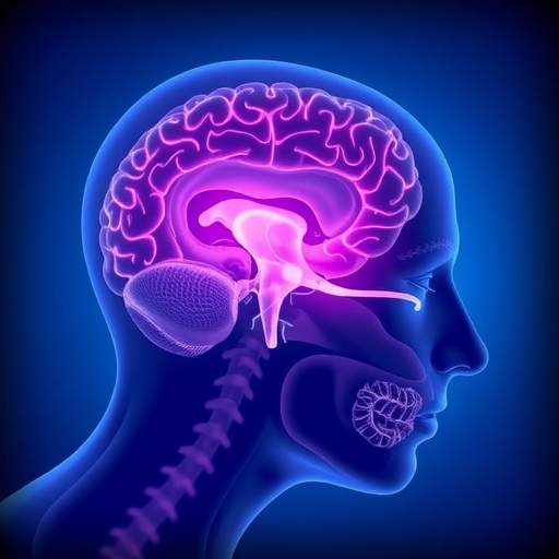
The glymphatic system, a brain-wide network responsible for the clearance of metabolic waste and interstitial solutes, has emerged as a pivotal player in maintaining neurological health. Its activity peaks during sleep, when cerebrospinal fluid (CSF) exchanges with interstitial fluid to facilitate the removal of neurotoxic proteins like alpha-synuclein and beta-amyloid. Impairment in this clearance pathway is increasingly recognized as a contributing factor in various neurodegenerative disorders, including Parkinson’s disease.
Utilizing advanced MRI techniques—specifically diffusion tensor imaging along the perivascular space (DTI-ALPS)—the study by Nepozitek and colleagues provides direct in vivo evidence of glymphatic dysfunction in PD patients. The DTI-ALPS method quantifies water diffusion along perivascular spaces, effectively serving as a biomarker for glymphatic efficiency. Reduced diffusivity metrics indicate a compromised glymphatic function, which correlates strongly with clinical symptoms.
.adsslot_pXRNzxuW0F{width:728px !important;height:90px !important;}
@media(max-width:1199px){ .adsslot_pXRNzxuW0F{width:468px !important;height:60px !important;}
}
@media(max-width:767px){ .adsslot_pXRNzxuW0F{width:320px !important;height:50px !important;}
}
ADVERTISEMENT
Notably, the research highlights a robust association between the intensity of obstructive sleep apnea symptoms and glymphatic dysfunction severity. Obstructive sleep apnea, characterized by repetitive upper airway obstruction during sleep, leads to intermittent hypoxia and fragmented sleep architecture. These disruptions likely impair the glymphatic clearance process, potentially fostering an environment conducive to neurodegenerative progression.
The study cohort comprised newly diagnosed Parkinson’s patients, a critical group for understanding early pathological mechanisms before extensive neurodegeneration sets in. The findings suggest that OSA severity could serve as an indicator or possibly a modifiable risk factor affecting glymphatic performance and, by extension, disease progression.
Pathophysiologically, the intersection of OSA and glymphatic dysfunction is thought to revolve around cerebrovascular dynamics and sleep quality. OSA-related hypoxia and intrathoracic pressure changes may disrupt perivascular fluid movement, compromising CSF flow along the glymphatic pathway. Moreover, the sleep fragmentation inherent in OSA reduces the duration of deep, slow-wave sleep—when glymphatic activity is most intense—thereby attenuating waste clearance.
By characterizing these mechanistic links, the study opens potential therapeutic avenues. Interventions targeting OSA—such as continuous positive airway pressure (CPAP) therapy—might restore glymphatic function, attenuate the accumulation of neurotoxic proteins, and slow Parkinson’s disease progression. This integrative approach could herald a paradigm shift in managing Parkinson’s, emphasizing early screening and treatment of sleep disorders as part of a holistic care strategy.
Technological advances underpinning this research are noteworthy. DTI-ALPS represents a non-invasive, sensitive, and replicable imaging modality capable of evaluating microstructural changes in glymphatic flow. Its application across a clinical setting may facilitate personalized monitoring of brain clearance functions, allowing clinicians to tailor interventions according to glymphatic integrity status.
Importantly, the study also raises questions about causality versus correlation. Is glymphatic dysfunction a consequence of OSA, a contributor to Parkinsonian neurodegeneration, or both? The bidirectional relationship merits further exploration through longitudinal and interventional trials to dissect how these systems influence each other over time.
Another intriguing implication relates to the timing of therapeutic interventions. Since glymphatic activity is tightly linked to sleep architecture, optimizing sleep quality early in the disease could maximize benefits, potentially delaying irreversible neuronal loss. Initiating OSA treatment promptly after Parkinson’s diagnosis might therefore yield neuroprotective effects beyond symptomatic relief.
Furthermore, this research complements emerging evidence spotlighting the role of vascular health in neurodegenerative diseases. Dysregulation in the brain’s clearance system may intersect with vascular impairments frequently observed in Parkinson’s patients, suggesting a multi-factorial cascade accelerating disease dynamics.
The study also underscores the importance of multidisciplinary collaboration, combining neurology, sleep medicine, and neuroimaging expertise to unravel complex disease networks. This integrative approach extends understanding beyond isolated mechanisms, fostering innovative diagnostics and personalized therapies.
At a cellular level, glymphatic failure impedes removal of misfolded proteins, exacerbating Lewy body formation—a hallmark of Parkinson’s pathology. The data suggest that OSA-induced hypoxia and disrupted sleep could heighten protein aggregation, fueling neuroinflammation and progressive motor and cognitive decline.
Moreover, the study prompts reevaluation of sleep disorders in neurodegenerative contexts. Rather than viewing OSA as a mere comorbidity, it highlights OSA as a potentially treatable driver of pathological processes. This reconceptualization encourages routine OSA screening in Parkinson’s patients, enhancing disease management protocols.
While these findings are promising, limitations exist. The cross-sectional design restricts causal inference, and larger, diverse cohorts are needed to validate and generalize results. Additionally, technological standardization of DTI-ALPS protocols will be essential for widespread clinical adoption.
Ultimately, this landmark research positions the glymphatic system and sleep-disordered breathing at the forefront of Parkinson’s disease investigation. It advocates for integrated diagnostic and therapeutic strategies that address the multifaceted nature of neurodegeneration. As the neuroimaging toolkit expands, coupling biological insight with clinical care may transform outcomes for millions affected globally.
As science continues to decode the enigmatic interplay between sleep, brain clearance, and neurodegeneration, studies like this pave the way toward innovative interventions. Bridging molecular mechanisms with clinical phenotypes, Nepozitek et al. illuminate new paths toward mitigating the heavy burden of Parkinson’s disease through targeted management of obstructive sleep apnea and preservation of glymphatic function.
Subject of Research: Glymphatic system dysfunction and its relationship to obstructive sleep apnea severity in newly diagnosed Parkinson’s disease patients.
Article Title: Glymphatic dysfunction evidenced by DTI-ALPS is related to obstructive sleep apnea intensity in newly diagnosed Parkinson’s disease.
Article References:
Nepozitek, J., Marecek, S., Rottova, V. et al. Glymphatic dysfunction evidenced by DTI-ALPS is related to obstructive sleep apnea intensity in newly diagnosed Parkinson’s disease. npj Parkinsons Dis. 11, 160 (2025). https://doi.org/10.1038/s41531-025-01018-8
Image Credits: AI Generated
Tags: advanced MRI techniques in neurologybrain clearance mechanisms in PDcerebrospinal fluid dynamics in sleepclinical symptoms of sleep apnea in PDDTI-ALPS imaging in Parkinson’s researchglymphatic system dysfunctionmetabolic waste clearance in the brainneurodegenerative disorders and sleepneurotoxic proteins and glymphatic healthobstructive sleep apnea in Parkinson’s diseaseParkinsonian pathology and sleep disordersperivascular space water diffusion imaging

