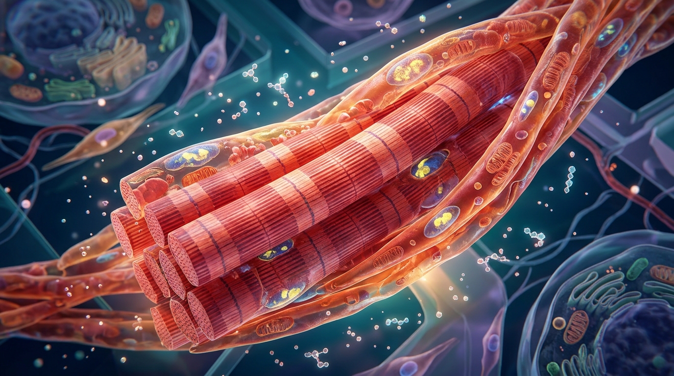
Yet the scale of that weight loss has carried a persistent worry: a measurable share of the mass disappearing from the body is lean tissue, including skeletal muscle. When primary human muscle cells were exposed directly to semaglutide or to GIP peptide, the cells showed no meaningful atrophy, no inflammatory shift, no mitochondrial damage and no impairment of the process by which muscle fibres form.
The research, published in GeroScience, was motivated by a specific clinical puzzle. Trials of semaglutide in adults with overweight or obesity have reported reductions in lean mass of up to roughly 9 per cent, while tirzepatide, a dual GLP-1 and GIP receptor agonist, has been associated with an approximately 11 per cent decline. Fat mass falls by more in absolute terms, so the ratio of fat to lean tissue improves, but the sheer loss of muscle has raised concerns because skeletal muscle governs mobility, physical function and metabolic health. Those concerns sharpen with age: sarcopenia, the progressive loss of muscle mass and function, affects an estimated 10 to 20 per cent of adults over 60 and is linked to falls, frailty, disability, hospitalisation and premature death.
The open question has been mechanism. Lean mass loss during rapid weight loss could simply be a by-product of the large caloric deficit and the reduced mechanical load that lighter body weight places on the skeleton, rather than a direct pharmacological effect of the drugs on muscle tissue. Their work is, to their knowledge, the first to test GLP-1 receptor and GIP receptor agonism directly on primary human skeletal muscle myotubes.
None of the participants were receiving incretin analogues or DPP-4 inhibitors prior to sample collection. Primary myoblasts were isolated from skeletal muscle of older adults with obesity undergoing orthopaedic surgery, grown in culture and differentiated into multinucleated myotubes.
The first question was whether the cells even carry the molecular targets for these drugs. The answer was asymmetric. GIP receptor mRNA was consistently detected in myotubes from all six donors, albeit at low levels, with delta-CT values between roughly 33 and 36 cycles. GLP-1 receptor mRNA, by contrast, was undetectable by PCR in every donor tested. Yet when the researchers used LUXendin645, a fluorescent probe that binds surface GLP-1 receptor protein, flow cytometry on myoblasts from three donors revealed a significant signal increase, with 51 to 66 per cent of cells classified as probe-positive. This discordance between undetectable mRNA and detectable surface protein is not unusual for very low-abundance receptors, and it suggests that any GLP-1 receptor expression in these cells is likely to be functionally limited.
With the receptor landscape mapped, the team exposed differentiated myotubes from six donors to semaglutide at 10 nanomolar, a concentration dosed to approximate total circulating plasma concentrations reported in clinical pharmacokinetic studies, or to GIP peptide at 5.02 micromolar, a dose several orders of magnitude above endogenous postprandial plasma GIP concentrations, selected deliberately to maximise sensitivity to a receptor-mediated effect given the absence of prior dosing literature. The authors note an important caveat on the semaglutide dose: because semaglutide is extensively bound to plasma albumin in vivo via an engineered fatty-acid albumin-binding moiety, and the albumin content of the differentiation medium is substantially lower than that of human plasma, the free, bioactive concentration to which cells were exposed in vitro is likely to exceed the free fraction present in circulation at the same nominal total concentration. After four hours of treatment, the expression of canonical atrophy genes, including MAFBX, MURF1 and FOXO3, and of inflammatory markers IL6 and TGFB, was unchanged relative to vehicle controls for both drugs.
A broader, unbiased sweep came from proteomics. Conditioned media collected after 24 hours of treatment were analysed using the Olink Reveal proximity extension assay panel, consisting of 1,034 proteins, with normalised protein expression values returned for 24 samples from six donors per condition. Principal component analysis showed that samples did not segregate by treatment: the first two principal components explained 40.3 per cent and 11.1 per cent of variance respectively, and the confidence ellipses of all groups overlapped extensively. After Benjamini-Hochberg correction across all 1,034 proteins, not a single protein reached statistical significance in either comparison, with the minimum adjusted p-value sitting at 1.0. A handful of proteins, including UBE2B, S100A12 and CRNN after GIP treatment and CD79B after semaglutide, cleared only nominal significance thresholds, and none of the six proteins most closely tied to muscle biology, FOXO1, MSTN, IGF1R, TGFB1, FOXO3 and IL6, showed significant changes.
Metabolism told a more nuanced story. Using the Seahorse XF Real-Time ATP Rate Assay in myotubes from four donors, the researchers found that semaglutide significantly reduced glycolytic proton efflux rate, glycolytic ATP production and total ATP production, with p-values of 0.0035, 0.0014 and 0.0187 respectively, while leaving oxygen consumption, mitochondrial ATP production, coupling efficiency and proton leak untouched. GIP had no effect on any of these bioenergetic measures. The authors interpret the semaglutide pattern as a modest suppression of glycolytic flux, mechanistically coherent with the reduced lactate, but they emphasise that the magnitude was small and not consistent with clinically meaningful muscle impairment. Whether the effect depends on canonical GLP-1 receptor signalling was not directly tested, for example by receptor antagonism, and remains open.
Morphology and myogenesis completed the picture. After 24 hours of treatment, neither drug changed myotube diameter, with p-values of 0.2824 for semaglutide and 0.5507 for GIP, while a TNF-alpha positive control confirmed the assay could detect genuine atrophy. Across an eight-day differentiation protocol with dosing every 48 hours, neither drug altered nuclear fusion index or myotube coverage, and MyoD and Myf5 expression were unaffected. One signal stood out: GIP reduced myogenin expression at day 4 by about 23 per cent, a statistically significant dip whose functional significance the authors call uncertain, since fusion and coverage at day 8 were normal. Intriguingly, GIP actually increased myotube thickness after eight days and reduced lactate dehydrogenase release relative to its vehicle, a combination the researchers read as a modest pro-hypertrophic or cytoprotective effect rather than harm.
The authors are careful about limits. The myotubes came from people with end-stage osteoarthritis, a condition marked by chronic inflammation and altered muscle metabolism, and the cultures were enriched rather than purified, so contaminating non-myogenic cells could contribute to the GLP-1 receptor protein signal. Most assays used six donors and may have been underpowered for subtle effects, the treatment windows cannot capture months of in vivo exposure, and the study used native GIP peptide rather than long-acting or dual agonists such as tirzepatide, so the findings should not be generalised to those engineered molecules without qualification. Even so, the overall conclusion is striking: direct GLP-1 and GIP receptor activation produced no major atrophic, inflammatory, morphological, myogenic or mitochondrial effects in these human muscle cells. That points the explanation for clinical lean mass loss away from the muscle cell itself and toward the caloric deficit and reduced mechanical loading that accompany weight loss, offering some reassurance as these drugs reach growing numbers of older adults at risk of sarcopenia.
Subject of Research: Direct effects of GLP-1 and GIP receptor agonists on primary human skeletal muscle myotubes
Article Title: GLP-1 and GIP receptor agonism does not directly drive skeletal muscle atrophy or impair myogenesis in primary human myotubes
Article References: Ditchfield, C., Macleod, M., Price, J. M. J., Davis, E. T., & Jones, S. W. (2026). GLP-1 and GIP receptor agonism does not directly drive skeletal muscle atrophy or impair myogenesis in primary human myotubes. GeroScience. https://doi.org/10.1007/s11357-026-02625-w
Image Credits: AI Generated
DOI: 10.1007/s11357-026-02625-w
Keywords: semaglutide, GIP, GLP-1 receptor, skeletal muscle, myotubes, lean mass loss, sarcopenia, incretin agonists, myogenesis, mitochondrial bioenergetics, glycolytic flux, obesity
News Source: Ophelia Keating. (November 4, 2026). GLP-1 and GIP drugs leave human muscle cells unharmed in lab study. Scienmag.