

• HOME
  • NEWS
  • EXPLORE
    • CAREER
      • Companies
      • Jobs
    • EVENTS
    • iGEM
      • News
      • Team
    • PHOTOS
    • VIDEO
    • WIKI
  • BLOG
  • COMMUNITY
    • FACEBOOK
    • INSTAGRAM
    • TWITTER
Sunday, August 16, 2026
BIOENGINEER.ORG
No Result
View All Result
  • Login
  • HOME
  • NEWS
  • EXPLORE
    • CAREER
      • Companies
      • Jobs
        • Lecturer
        • PhD Studentship
        • Postdoc
        • Research Assistant
    • EVENTS
    • iGEM
      • News
      • Team
    • PHOTOS
    • VIDEO
    • WIKI
  • BLOG
  • COMMUNITY
    • FACEBOOK
    • INSTAGRAM
    • TWITTER
  • HOME
  • NEWS
  • EXPLORE
    • CAREER
      • Companies
      • Jobs
        • Lecturer
        • PhD Studentship
        • Postdoc
        • Research Assistant
    • EVENTS
    • iGEM
      • News
      • Team
    • PHOTOS
    • VIDEO
    • WIKI
  • BLOG
  • COMMUNITY
    • FACEBOOK
    • INSTAGRAM
    • TWITTER
No Result
View All Result
Bioengineer.org
No Result
View All Result
Home NEWS Science News Biology

Forces of Push and Pull Shape How Blood Flows Through Vessels

Bioengineer by Bioengineer
July 28, 2026
in Biology
Reading Time: 2 mins read
0
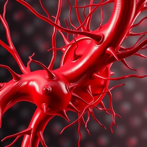
Forces of Push and Pull Shape How Blood Flows Through Vessels
Share on FacebookShare on TwitterShare on LinkedinShare on RedditShare on Telegram

Blood vessels must form in a way that is both precise and reliable, so that oxygen and nutrients can reach every part of the body. During development, endothelial cells organize into a branched network of hollow tubes, creating a continuous lumen that later allows blood to flow. Yet how neighboring vessel segments coordinate their movement and connect into an unbroken channel has remained only partially understood.

Using high-resolution live imaging in zebrafish, researchers from the University of Basel—led by Professor Markus Affolter and Dr. Heinz-Georg Belting—mapped the process with unprecedented detail. Their work, published in eLife, reveals the full sequence by which adjacent lumina merge to generate mature, connected blood vessels.

The study focuses on the junctions that keep endothelial cells attached to one another during tube formation. These cell-cell contacts must be stable enough to preserve the integrity of the vessel wall, while also remaining adaptable to permit rearrangements of the cells.

A key player in this remodeling is junction-based lamellipodia (JBL), specialized membrane protrusions that drive endothelial cell repositioning. The new findings explain the mechanical logic behind how these JBL structures enable cells to extend, meet, and link.

The process begins when a JBL protrusion forms at the tip of an endothelial cell, producing a pushing force that advances the cell front. At the protrusion’s tip, the junction acts like a molecular anchor, attaching to a neighboring cell and stabilizing the growing interface between segments.

Then pulling forces take over, drawing the rear of the cell forward and gradually elongating the cell. In repeated push-and-pull cycles, cells behave in an inchworm-like manner, progressively extending the lumen until separate segments fuse into a continuous vessel.

The researchers report that VE-cadherin, a major junction molecule, plays two distinct roles: it provides “glue” for adhesion and vessel-wall stability, and it actively supports cell movement. Notably, endothelial cells appear both robust and highly plastic during the same developmental steps.

Beyond clarifying vascular development, the work suggests general principles for how tissues could be patterned around working blood supply. Because many organs develop in close coordination with vasculature, improved understanding may translate into better strategies for engineering vascularized tissues.

In the long term, these insights could help create organoids and laboratory-grown tissues that include functional blood vessels—an essential step toward making regenerative medicine more faithful to natural development.

Subject of Research: Endothelial cell mechanics and junction-mediated vascular tube formation
Article Title: Junctional and Actomyosin Dynamics Drive Endothelial Cell Rearrangements during Vascular Tube Formation.
Web References: http://dx.doi.org/10.7554/eLife.109264.2
References: eLife, 10.7554/eLife.109264.2
Image Credits: Etienne Schmelzer, Biozentrum, University of Basel

Tags: blood vessel formationblood vessel morphogenesis mechanismscell-cell adhesion in blood vessel developmentendothelial cell junction remodelingendothelial cell migration and connectionforces driving blood vessel fusionhigh-resolution imaging of vascular tissuesjunction-based lamellipodia in blood vesselslive imaging of blood vessel developmentvascular network patterningvessel lumen formationzebrafish models in vascular research
Share12Tweet7Share2ShareShareShare1

Related Posts

Strubbelig–NHL3 Receptor Complex Helps Arabidopsis Respond to Cellulose Deficiency

Strubbelig–NHL3 Receptor Complex Helps Arabidopsis Respond to Cellulose Deficiency

August 15, 2026
Bombyx mori Satellitome Analysis Reveals Evolutionary Stability, Dispersed Chromosomal Organization, Transposon Origins

Bombyx mori Satellitome Analysis Reveals Evolutionary Stability, Dispersed Chromosomal Organization, Transposon Origins

August 15, 2026

Synthetic pyrenoid reconstruction reveals EPYC1-driven carbon concentration mechanisms and evolution

August 15, 2026

Endophytic Flavobacterium boosts root hairs and drought tolerance through ERF–CEP5 signaling

August 15, 2026

POPULAR NEWS

  • KAIST develops semiconductor neuron that harnesses noise to selectively process signals

    29 shares
    Share 12 Tweet 7
  • Imagining natural and extra robotic thumbs together strengthens kinesthetic sensorimotor networks

    29 shares
    Share 12 Tweet 7
  • PARP1 Drives Neuropathic Pain Through GPX4-Dependent Ferroptosis in Injured Mice’s Sensory Neurons

    29 shares
    Share 12 Tweet 7
  • Strubbelig–NHL3 Receptor Complex Helps Arabidopsis Respond to Cellulose Deficiency

    29 shares
    Share 12 Tweet 7

About

We bring you the latest biotechnology news from best research centers and universities around the world. Check our website.

Follow us

Recent News

KAIST develops semiconductor neuron that harnesses noise to selectively process signals

Imagining natural and extra robotic thumbs together strengthens kinesthetic sensorimotor networks

PARP1 Drives Neuropathic Pain Through GPX4-Dependent Ferroptosis in Injured Mice’s Sensory Neurons

Subscribe to Blog via Email

Success! An email was just sent to confirm your subscription. Please find the email now and click 'Confirm' to start subscribing.

Join 85 other subscribers
  • Contact Us

Bioengineer.org © Copyright 2023 All Rights Reserved.

Welcome Back!

Login to your account below

Forgotten Password?

Retrieve your password

Please enter your username or email address to reset your password.

Log In
No Result
View All Result
  • Homepages
    • Home Page 1
    • Home Page 2
  • News
  • National
  • Business
  • Health
  • Lifestyle
  • Science

Bioengineer.org © Copyright 2023 All Rights Reserved.