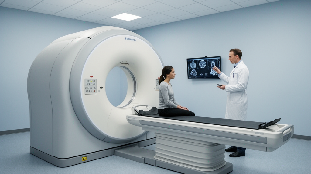
Patients treated for early-stage kidney cancer are undergoing more follow-up CT scans than guidelines recommend, and most of those scans fail to detect a return of the disease, according to a retrospective cohort study published in European Radiology Abdomen. The findings, drawn from 228 patients treated at a Dutch tertiary center between January 2016 and September 2022, add real-world patient-level data to a growing debate about whether intensive imaging surveillance after curative treatment for non-metastatic renal cell carcinoma is justified.
Renal cell carcinoma accounts for roughly 3 percent of malignancies worldwide, and its incidence is rising by about 2 percent annually, largely because tumors are increasingly discovered incidentally on imaging done for other reasons. About one-third of patients diagnosed with non-metastatic disease will eventually experience a local recurrence or distant metastases after curative treatment. Follow-up after surgery or ablation relies predominantly on CT imaging, with schedules stratified by risk according to guidelines from the European Association of Urology (EAU), the National Comprehensive Cancer Network, or the American Urological Association. The 2025 EAU guideline recommends chest-abdomen CT at intervals determined by risk category, based on the Leibovich score, pathological TNM staging, and ISUP grade, while acknowledging that the evidence base for these recommendations is limited.
Eligible patients were adults with histologically confirmed, de novo non-metastatic renal cell carcinoma who underwent curative-intent surgery, either radical or partial nephrectomy, or tumor ablation by cryoablation, microwave ablation, or radiofrequency ablation. Patients with primary metastatic disease, hereditary RCC syndromes, or multiple unilateral or bilateral tumors were excluded. Of 411 eligible consecutive patients, 228 were included; the rest were excluded for lack of histopathological proof of RCC, absence of active treatment, multifocal tumors, or hereditary syndromes.
Patients were classified into Leibovich risk groups: 127 as low risk, 63 as intermediate risk, and 38 as high risk. For non-clear-cell tumors, risk classification was based on pathological TNM stage and ISUP grade. The mean age of the cohort was 66.9 years, and the mean follow-up duration across all risk groups was 49.1 months, with data collected until June 26, 2025. All CT findings were based on the original radiological reports, and no independent image re-review was performed. Only chest-abdomen CTs performed within the urological follow-up setting were counted; scans requested by other departments for unrelated indications were excluded.
Compared with the 2025 EAU recommendations, which prescribe four CTs per five years for low-risk patients, six for intermediate risk, and eight for high risk, the cohort was imaged more often than advised. Adjusted for each patient’s actual follow-up duration, the guideline-expected numbers of scans were 2.6, 3.8, and 4.9 for the low-, intermediate-, and high-risk groups, respectively. The observed means were 4.3, 4.9, and 5.0. Among patients with at least five years of follow-up, the averages were 6.5, 8.1, and 8.1 scans, against recommendations of 4, 6, and 8. Low-risk patients thus underwent a median of six follow-up CT scans, exceeding even the higher frequencies recommended by previous versions of the EAU guideline. In the high-risk group, the observed number did not exceed the recommended count, though most of those patients were still under follow-up when the database closed.
In total, 1,046 CT scans were performed across the cohort. Abnormal findings were common: 66.9 percent of low-risk, 68.3 percent of intermediate-risk, and 78.9 percent of high-risk patients showed at least one abnormal finding during follow-up. But abnormal did not mean cancerous. Only a subset of abnormalities were potentially RCC-related, between 43.3 and 60.5 percent depending on risk group, and confirmed recurrence or metastases, defined as pathologically verified lesions consistent with RCC on histological examination, were found in just 2.4 percent of low-risk patients, 15.8 percent of intermediate-risk patients, and 18.5 percent of high-risk patients. In the low-risk group, confirmed local recurrence occurred in two of 127 patients and confirmed metastatic disease in one patient with a liver lesion; no patient developed confirmed lung metastases.
The cascade from suspicious image to confirmed disease was strikingly inefficient. Among low-risk patients whose CT findings triggered a change in management, 29 of 32, or 90.6 percent, had no confirmed recurrence or metastases. Most of those suspicious findings resolved spontaneously or remained stable over years of observation. In the intermediate-risk group, 11 of 21 patients with management changes, 52.4 percent, had no confirmed disease; in the high-risk group, the figure was 10 of 17, or 58.8 percent. A small but clinically relevant subset of these work-ups did yield something else: a new primary malignancy was diagnosed in five low-risk, four intermediate-risk, and one high-risk patient, a potential benefit of surveillance that extends beyond kidney cancer detection, though the authors note that whether this justifies the current imaging intensity remains an open question.
Incidental findings unrelated to RCC added a further layer of diagnostic noise, recorded in 23.6 percent of low-risk, 19.0 percent of intermediate-risk, and 18.4 percent of high-risk patients. These included cysts, vascular abnormalities, hepatobiliary findings, and benign skeletal lesions. Suspicious pulmonary nodules, which are common and usually unrelated to RCC, appeared in 7.9 percent of low-risk patients, none of whom developed confirmed lung metastases. Current guidelines lack specific recommendations for managing such nodules, leaving decisions to individual physicians and potentially prompting additional imaging. The authors note that the lungs are the most frequent site of RCC metastases, at 45 to 75 percent, which makes nodules a recurring source of concern even when they turn out to be benign.
The study’s authors argue that the low cancer-specific yield, particularly among low-risk patients, warrants a critical appraisal of current follow-up intensity. Their conclusions align with earlier evidence: an analysis of the large RECUR database by Dabestani and colleagues showed that cross-sectional imaging for detecting recurrences after surgery for localized RCC did not improve overall survival, and a systematic review of 20 studies reached a similar conclusion. Among patients in the new cohort with confirmed recurrence or metastases, the median time from treatment to the confirmed event was 29 months in the low-risk group, 19.5 months in the intermediate-risk group, and 15 months in the high-risk group. The authors also point to work suggesting that in low-risk patients aged 80 or older, or with a Charlson Comorbidity Index of 2 or higher, the risk of death from causes other than RCC exceeds the risk of recurrence as early as six months after surgery, supporting an individualized approach to surveillance.
The study has limitations that the authors acknowledge. Confirmed recurrence relied on pathological verification, which may underestimate the true number of recurrences, although in this cohort all suspicious findings were either pathologically confirmed, resolved spontaneously, or attributed to a second primary malignancy. Patients were treated at a university medical center, which may limit generalizability, and the high-risk group was small at 38 patients. The retrospective design introduces the risk of information bias and precludes causal inference, and a formal distinction between scheduled surveillance CTs and symptom-driven scans was not systematically made. Not all patients completed the full five-year follow-up period, so late recurrences may have been missed, and quality-of-life aspects, including the anxiety that can accompany frequent scanning, were not assessed. The cohort also spanned 2016 to 2025 while the benchmark is the 2025 EAU guideline, though the authors note that recommended frequencies have not changed substantially between guideline versions. Emerging tools such as urinary glycosaminoglycan biomarkers, which showed high sensitivity for detecting clear-cell RCC recurrence in one study, may eventually help personalize follow-up, but prospective trials are needed. For now, the authors conclude that prospective evaluation of risk-adapted surveillance de-escalation is warranted, and that any reduction in scan frequency should be discussed with patients through shared decision-making.
Subject of Research: Diagnostic yield and surveillance burden of follow-up CT imaging after curative treatment for non-metastatic renal cell carcinoma
Article Title: Surveillance burden of CT imaging during follow-up for patients after curative treatment for non-metastatic renal cell carcinoma
Article References: Reijerink, M. A. A., Zondervan, P. J., van Delden, O. M., Bipat, S., & Stoker, J. (2026). Surveillance burden of CT imaging during follow-up for patients after curative treatment for non-metastatic renal cell carcinoma. European Radiology Abdomen, 1(1), Article 5. https://doi.org/10.1007/s44501-026-00007-3
Image Credits: AI Generated
DOI: 10.1007/s44501-026-00007-3
Keywords: renal cell carcinoma, CT surveillance, follow-up imaging, EAU guidelines, diagnostic yield, incidental findings, recurrence detection, Leibovich risk score, overdiagnosis, radiation exposure, kidney cancer, surveillance de-escalation
News Source: Nathaniel Bowman. (October 28, 2026). Follow-up CT Scans After Kidney Cancer Cure Find Little, Study Suggests. Scienmag.