


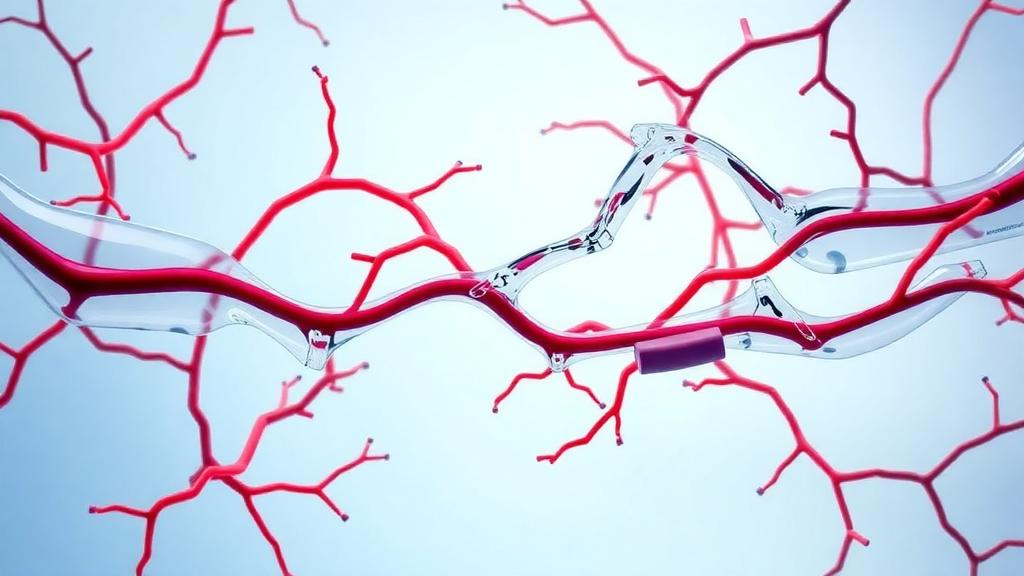
When a patient is in shock, doctors reach for intravenous fluids with a simple hope: push more volume in, push more blood out of the heart, and rescue the tissues starved of oxygen. But a new review published in Annals of Intensive Care argues that this chain of logic breaks down at the smallest scale of the circulation, where the real business of oxygen delivery takes place.
The review’s central finding is that fluid administration probably improves sublingual microcirculation when tissue perfusion is altered and cardiac output actually rises. That second condition matters more than it might seem. Shock states are characterized by a partial loss of coherence between systemic hemodynamics and the microcirculation, meaning that a boost in cardiac output does not guarantee improvements in tissue perfusion. In septic shock, in particular, the outcome of patients is not related to the magnitude of cardiac output but to tissue perfusion itself. Nonsurvivors of septic shock exhibit lower perfused capillary density, lower proportions of perfused microvessels, and lower red blood cell velocity than survivors, along with increased flow heterogeneity, and these derangements appear regardless of whether the patient’s heart is pumping in a hyperdynamic or normal fashion.
Why would more cardiac output fail to translate into better microvascular flow? The review lays out several mechanisms by which fluids can become a double-edged sword. Fluid administration carries risks of hemodilution and tissue edema, both of which can dampen microcirculation. Tissue edema can decrease vascular density and increase the oxygen diffusion distance from capillaries to mitochondria. Rising venous pressure may also compromise microvascular flow, although the physiology here is contested and depends on whether one considers tissue perfusion to depend on a vascular waterfall effect that renders flow independent of downstream pressures. Fluids can even provoke compartment syndrome in capsulated organs such as the kidney.
Hemodilution emerges as one of the most instructive examples of the disconnect between the macrocirculation and the microcirculation. In sheep exposed to stepwise hemodilution, cardiac output climbed progressively even as total and perfused vascular densities, the proportion of perfused vessels, and red blood cell velocity all fell in intestinal and sublingual mucosa. All microcirculatory variables were strongly correlated with hemoglobin level. Moderate hemodilution, up to roughly 15 percent reduction in hematocrit, was linked to improved microcirculation, but more extreme dilution produced progressive and severe microvascular derangements paradoxically accompanied by high cardiac output. The review’s authors describe extreme hemodilution as the most paradigmatic condition of dissociation between systemic hemodynamics and microcirculation. The mechanism appears to involve reduced blood viscosity, which lowers shear stress on the vascular wall and decreases production of nitric oxide, causing vasoconstriction and loss of perfused vessels.
Against this backdrop of physiological caution, the clinical evidence offers a qualified message of hope. The review identified eight observational and four controlled trials carried out in critically ill and surgical patients. Most showed that fluid resuscitation can improve microcirculation, especially in the presence of fluid responsiveness and tissue hypoperfusion. In patients with sepsis and high values of respiratory pulse pressure variation, either passive leg raising or volume expansion with saline or hydroxyethyl starch increased sublingual microcirculatory perfusion. A bolus of hydroxyethyl starch produced favorable effects on most sublingual microcirculatory variables in septic patients who simultaneously responded with increased stroke volume, whereas such effects were not found in nonresponders. In surgical patients, alterations in sublingual microcirculation were linked to preload dependence and were corrected after a fluid challenge. The effects of a fluid challenge on sublingual red blood cell velocity and perfused vascular density were strongly correlated with the basal state of the microcirculation and the magnitude of fluid responsiveness, meaning the benefits were most pronounced in patients with the deepest hypoperfusion and the largest increases in cardiac output.
Timing also appears to matter. In the early phase of sepsis, the administration of 1,000 milliliters of Ringer lactate or 400 milliliters of 4 percent albumin increased the sublingual proportion of perfused vessels and the perfused vascular density, but this effect was lost in later stages. Notably, no relationship between these microvascular effects and changes in cardiac index or mean arterial pressure was observed. Not every study was positive, however. A small randomized controlled trial in abdominal surgery patients found no microcirculatory benefit despite higher fluid volumes and increased cardiac output, although baseline microcirculatory variables were only slightly altered in both groups, leaving little room for improvement.
The thorniest question is which fluid to choose, and here the review delivers a verdict of unresolved uncertainty. Concerning the controlled trials that compared different solutions, one study failed to show benefits of hypertonic over isotonic hydroxyethyl starch, while another found improved microcirculation after early goal-directed therapy with hydroxyethyl starch compared with 0.9 percent sodium chloride. Since both studies included small samples, the results are inconclusive. A third trial, which recruited 100 septic patients and concluded that albumin was superior to a balanced solution, is flawed by methodological problems: the authors concluded that 20 percent albumin improves microcirculation, but this is not clearly supported by the results, since microvascular changes were minor and significant time-by-group interactions were not shown for any microcirculatory variable. The clinical usefulness of albumin for recruiting microcirculation in septic patients therefore remains uncertain.
Experimental studies add further layers of controversy. Some studies suggesting benefits of albumin, hydroxyethyl starch, and high-viscosity solutions could not be properly replicated in patients. Hydroxyethyl starch did outperform saline in rabbit and septic animal models, in one case decreasing microvascular leukocyte adherence and increasing functional capillary density despite similar systemic cardiovascular effects, but randomized controlled trials have shown that hydroxyethyl starch is not superior to crystalloids for hemodynamic resuscitation, has toxic effects, and increases mortality of septic patients, so it should not be used in critically ill patients. Hypertonic solutions showed favorable microvascular actions in some experimental work but contradictory evidence elsewhere, and in the one controlled study in septic shock patients they produced no differences in sublingual microcirculation compared with isotonic fluid. The superiority of balanced crystalloids over 0.9 percent sodium chloride was not consistently demonstrated in basic research either, and no clinical study has compared balanced crystalloid and saline on sublingual microcirculation. Albumin-derived molecules such as polyethylene glycol-conjugated albumin and a protein cocktail of human serum albumin, transferrin, haptoglobin, and hemopexin showed promising microcirculatory effects in experimental hemorrhagic shock, but none of these molecules has been clinically evaluated.
The review also raises a fundamental question about the sublingual window itself. The sublingual mucosa is easily accessible for videomicroscopic evaluation, and videos can be acquired with minimal patient discomfort, but its relationship to other microvascular beds is controversial. In septic patients, fluid challenges recruited sublingual but not stoma mucosa, and patient outcomes were linked to intestinal but not sublingual microcirculation. Contrast-enhanced ultrasound showed decreased renal cortical perfusion that was not adequately reflected by changes in sublingual microcirculation. Septic patients may therefore exhibit intestinal and renal microvascular disorders with concomitantly normal sublingual perfusion, a caveat that tempers any enthusiasm for guiding therapy by the tongue alone.
Compounding all of this, the review identifies severe methodological weaknesses in some of the studies it analyzed. Some microcirculatory videos were analyzed by unreliable methodologies, including point of care assessment with poor reproducibility and the AVA 4.3 software, which has never been validated and has been shown to produce wide limits of agreement and values outside the physiological range. Videos posted as typical examples in the supplementary material of some studies show severe compression artifacts and should have been deleted from the analysis. These pitfalls, the authors argue, preclude relevant conclusions about the effects of fluids on sublingual microcirculation. The bottom line of the review is measured: fluid administration probably improves sublingual microcirculation when tissue perfusion is altered and cardiac output increases, but the superiority of any solution for this purpose has not been clearly demonstrated, and high-quality studies are needed to clarify the effects of different fluids on the microcirculation that ultimately decides whether tissues live or die.
Subject of Research: Effects of fluid resuscitation on sublingual microcirculation in critically ill patients
Article Title: Effects of fluids on sublingual microcirculation: a point of view review
Article References: Dubin, A. (2025). Effects of fluids on sublingual microcirculation: a point of view review. Annals of Intensive Care, 15(1), Article 178. https://doi.org/10.1186/s13613-025-01607-z
Image Credits: AI Generated
DOI: 10.1186/s13613-025-01607-z
Keywords: microcirculation, fluid resuscitation, shock, sepsis, sublingual videomicroscopy, hemodilution, albumin, hydroxyethyl starch, crystalloids, colloids, intensive care, hemodynamics
News Source: Ophelia Keating. (October 29, 2026). Fluids Can Rescue the Microcirculation in Shock, but No Solution Wins Yet. Scienmag.



