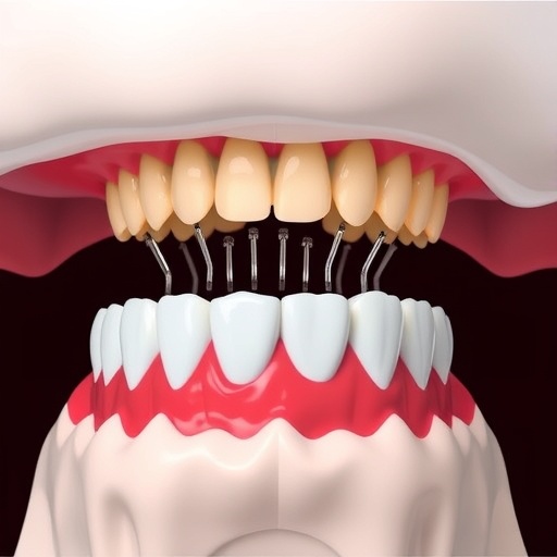
In recent years, the cellular process known as ferroptosis—an iron-dependent form of regulated cell death characterized by the accumulation of lipid peroxides—has drawn significant attention within the biomedical field. Emerging evidence has established ferroptosis as a pivotal factor in various inflammatory diseases. One condition increasingly linked to ferroptosis is periodontitis, a chronic inflammatory disorder that progressively destroys the connective tissues supporting the teeth. Despite its prevalence and impact on oral health worldwide, the precise role of ferroptosis in the pathogenesis and progression of periodontitis remains an intriguing frontier, demanding deeper scientific exploration.
Periodontitis stems from microbial plaque accumulation, primarily driven by anaerobic bacteria within periodontal pockets. These microbial colonies trigger a sustained inflammatory response, contributing to tissue degradation. Recent mechanistic studies have revealed multiple interlinked pathways through which ferroptosis accelerates periodontal tissue damage. Central to these mechanisms is the iron overload observed in inflamed periodontal tissues. A high-iron environment, exacerbated by both systemic iron metabolic disorders and local microbial activity, creates conditions that facilitate the initiation and propagation of ferroptosis within periodontal cells.
Multiple systemic disorders known for dysregulated iron metabolism, such as genetic hemochromatosis and sickle cell anemia, heighten the severity of periodontitis by contributing to excessive iron deposition in periodontal tissues. When microbial invasion occurs—especially within 12 hours post-infection—free iron levels elevate sharply, spurring biofilm maturation and bacterial proliferation. Notably, periodontal pathogens like Porphyromonas gingivalis and Prevotella intermedia possess strategies to hijack host iron-binding proteins, degrading them to release free iron. This not only supports microbial growth but also prompts localized iron overload, a critical driver of ferroptotic cascades.
.adsslot_Y1sntgdkyi{width:728px !important;height:90px !important;}
@media(max-width:1199px){ .adsslot_Y1sntgdkyi{width:468px !important;height:60px !important;}
}
@media(max-width:767px){ .adsslot_Y1sntgdkyi{width:320px !important;height:50px !important;}
}
ADVERTISEMENT
Hypoxia within periodontal pockets further compounds this iron dysregulation. The predominance of anaerobic bacteria fosters a low-oxygen microenvironment, activating hypoxia-inducible factor-1α (HIF-1α) in periodontal cells. This transcription factor indirectly intensifies iron accumulation by upregulating transferrin receptor (TFR) and heme oxygenase-1 (HO-1), facilitating heightened intracellular iron levels. The ensuing iron surplus predisposes periodontal cells to ferroptosis, thereby aggravating inflammatory tissue destruction.
Beyond iron overload, lipid peroxidation serves as a hallmark of ferroptotic cell death and is deeply implicated in periodontitis pathology. Studies have consistently shown significantly elevated lipid peroxide concentrations in saliva and gingival crevicular fluid of affected individuals. Alterations in key trace metal ions—Fe²⁺, Na⁺, Mg²⁺, Ca²⁺—in these fluids markedly influence redox homeostasis and exacerbate oxidative stress within the periodontal milieu. Elevated Fe²⁺ catalyzes the generation of reactive oxygen species (ROS), inducing lipid peroxidation that destabilizes cellular membranes.
The perturbation of the cellular antioxidant defense system further precipitates ferroptosis in periodontal tissues. Increased sodium ion concentrations correlate with the suppression of the system Xc⁻–GPX4 axis, limiting cystine import and glutathione (GSH) synthesis. As GSH depletion undermines glutathione peroxidase 4 (GPX4) activity, cells lose their ability to detoxify lipid peroxides, visible through rising malondialdehyde (MDA) levels, a lipid peroxidation byproduct and a sensitive ferroptosis biomarker. Simultaneously, reduced magnesium availability curtails ATP production within macrophages, impairing antioxidant enzyme synthesis and weakening cellular resilience to oxidative stress.
Calcium ions also play a pivotal role by activating phospholipase A2 (PLA2), initiating phospholipid degradation that liberates arachidonic acid (AA). Subsequently, lipoxygenases (LOX) convert AA into cytotoxic lipid peroxides, intensifying ferroptosis induction. Neutrophils infiltrating periodontal sites generate abundant ROS via NADPH oxidase-mediated respiratory bursts, creating a self-amplifying cycle of oxidative damage and ferroptosis acceleration.
Ferritinophagy, the selective autophagic degradation of ferritin leading to intracellular iron release, represents another critical mechanism implicated in periodontitis-associated ferroptosis. The inflamed periodontal microenvironment appears to stimulate ferritinophagy, resulting in abnormal iron deposition within periodontal ligament fibroblasts and other resident cells. Hypoxia-inducible factors, modulated by NF-κB signaling, govern the expression of iron homeostasis proteins, including ferritin, highlighting their importance in this context. Concurrently, T cells infiltrating periodontal tissue may contribute to ferritin presence, although direct evidence of their role remains nascent.
Dysbiotic shifts in oral microbiota, dominated by anaerobic species such as P. gingivalis, Actinomyces, and Fusobacterium nucleatum, promote the production of short-chain fatty acids (SCFAs) like butyrate. Butyrate particularly enhances nuclear receptor coactivator 4 (NCOA4) expression, triggering ferritinophagy and liberating free iron. This cascade depletes cellular GSH and GPX4 reserves, upregulates acyl-CoA synthetase long-chain family member 4 (ACSL4), and intensifies lipid peroxidation. Meanwhile, periodontal pathogens exploit iron derived from ferritin degradation to sustain their metabolic needs and virulence, thereby perpetuating the inflammatory environment.
At the molecular signaling level, ferroptosis in periodontal cells is orchestrated by complex and multifaceted pathways intertwining with inflammation and oxidative stress response networks. The transforming growth factor-beta (TGF-β) pathway stands out as a significant ferroptosis facilitator. Inflammatory stimuli drive periodontal cells to overexpress TGF-β, which activates the TNF receptor 1 (TNFR1) and nuclear factor kappa B (NF-κB) signaling, upregulating iron transport proteins such as DMT1 and fostering Fe²⁺ accumulation intracellularly. TGF-β1 mediates ferroptosis through Smad3 signaling by repressing solute carrier family 7 member 11 (SLC7A11), a cystine-glutamate antiporter pivotal for antioxidant defense.
Moreover, TGF-β synergizes with interleukin-6 (IL-6) to activate the JAK/STAT3 pathway, which enhances hepcidin expression—a systemic iron regulator that enforces intracellular iron retention. This cytokine milieu also modulates macrophage polarization toward the M2 phenotype, associated with increased expression of iron uptake receptors like CD163. These macrophages contribute to iron sequestration within periodontal tissue, exacerbating ferroptosis susceptibility.
Conversely, the nuclear factor erythroid 2-related factor 2 (Nrf2) pathway serves as a vital cellular defense mechanism countering ferroptosis. As the master regulator of antioxidant responses, Nrf2 controls the transcription of genes coding for antioxidant enzymes including GPX4 and ferritin. In periodontitis, Nrf2 signaling is notably suppressed, weakening redox homeostasis and facilitating lipid peroxide accumulation. Activation of Nrf2 has been demonstrated to restore antioxidant capacity, lower lipid peroxide burden, and inhibit ferroptotic processes, thus offering promising therapeutic avenues for periodontal inflammation management.
Another pivotal player is the tumor suppressor protein p53—a dual-function regulator with context-dependent influences on ferroptosis. Under hypoxic and inflammatory stimuli characteristic of periodontitis, p53 expression is elevated and correlates with disease severity. Intriguingly, p53 can potentiate ferroptosis by repressing SLC7A11 and inducing spermidine/spermine N1-acetyltransferase 1 (SAT1), thereby enhancing ALOX15-mediated lipid peroxidation. Conversely, p53 exhibits inhibitory effects on ferroptosis through modulation of dipeptidyl peptidase-4 (DPP4) activity and induction of CDKN1A/p21, which mitigate oxidative stress. However, the precise molecular interplay between p53 and ferroptotic pathways in periodontal tissues remains incompletely understood and represents an active field of research.
Cumulatively, these insights delineate a complex network whereby microbial activity, iron metabolism dysregulation, oxidative stress, and autophagic pathways converge to activate ferroptosis, fueling periodontal tissue destruction. The intersection of local environmental factors—such as hypoxia, altered metal ion concentrations, and microbial metabolites—and intrinsic cellular signaling cascades creates a fertile ground for ferroptosis-mediated cellular demise in periodontitis.
Understanding the nuanced mechanisms underlying ferroptosis in periodontal disease not only advances our comprehension of its pathogenesis but also illuminates novel molecular targets for therapeutic intervention. Strategies aimed at modulating iron overload, enhancing antioxidant defenses via Nrf2 activation, or inhibiting key ferroptosis drivers such as TGF-β and ferritinophagy hold promise for curbing periodontal inflammation and halting tissue degradation.
The research community continues to investigate how periodontal pathogens and their metabolic byproducts impact ferroptosis directly, particularly regarding the PUFA/LOX axis and lipid peroxide generation. Additionally, clarifying the contributions of immune components like T cells in ferritin dynamics and defining the precise roles of p53 modulatory functions in oral tissues will be critical for translating these findings into clinical applications.
Periodontitis represents a multifactorial disease with systemic implications, and ferroptosis emerges as a central pathological process intertwining local infection, immune response, and metabolic dysregulation. As studies unfold, harnessing ferroptosis modulation may revolutionize periodontal therapy, transforming how clinicians approach this widespread inflammatory disease and potentially mitigating its substantial morbidity on oral and overall health.
Subject of Research:
The role and mechanisms of ferroptosis in the progression and pathogenesis of periodontitis.
Article Title:
Ferroptosis in periodontitis: mechanisms, impacts, and systemic connections.
Article References:
Guan, P., Ruan, Q., Li, J. et al. Ferroptosis in periodontitis: mechanisms, impacts, and systemic connections. Cell Death Discov. 11, 283 (2025). https://doi.org/10.1038/s41420-025-02550-5
Image Credits:
AI Generated
Tags: anaerobic bacteria in periodontal diseasechronic inflammatory disordersferroptosis in periodontitisinflammatory diseases and ferroptosisiron metabolism disordersiron overload in oral healthlipid peroxides and cell deathmechanisms of ferroptosismicrobial influence on periodontitisperiodontal tissue damageperiodontitis pathogenesissystemic iron dysregulation


