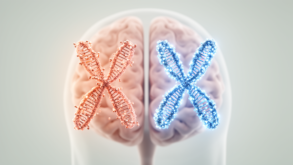
The female and male X chromosomes are not methylated the same way in the human brain, according to a new chromosome-wide analysis of DNA methylation published in the journal Epigenetics & Chromatin. Drawing on 89 whole-genome methylomes generated from human neurons and oligodendrocytes, researchers report that the combined methylation of the two female X chromosomes is globally lower than that of the single male X chromosome, a pattern that holds across the chromosome as a whole rather than being confined to a few isolated regions.
DNA methylation is a key epigenetic mechanism involved in the regulation of sex chromosomes, yet the authors of the study note that our understanding of X chromosome methylation has lagged behind that of the autosomes. Many DNA methylation studies have excluded X chromosomes altogether, and widely used methylation assays interrogate biased subsets of CpG sites rather than assessing CpGs across the whole chromosome. The new work set out to close that gap by using an unbiased, chromosome-wide dataset.
The study, led by Yong Hwee Eddie Loh, Robert Morgan, Devika Singh, Isabel Mendizabal and Soojin V. Yi, examined methylation in neurons and oligodendrocytes, two cell types of the human brain. Because the analysis covered whole genomes rather than selected CpG panels, the researchers were able to compare methylation levels between the female X chromosomes and the male X chromosome across the entire chromosome, as well as against autosomal methylation levels.
The headline result is that the combined methylation of the female X chromosomes is globally reduced, or hypomethylated, across the chromosome compared with the male X and with autosomes. This chromosome-wide reduction was corroborated by an analysis of differentially methylated regions distributed across the whole X chromosome, which also revealed many regions that differ between the two sexes and between the two examined cell types.
At the level of promoters, the picture is more heterogeneous. While many promoters are more highly methylated, or hypermethylated, on the female X than on the male X, a substantial number of promoters show the opposite pattern, with lower methylation in females. This mixed landscape contrasts with the pervasive chromosome-wide reduction seen when methylation is considered across the X chromosome as a whole.
One of the most striking findings concerns genes with higher expression in females, which the researchers used as a proxy for genes that escape X chromosome inactivation. The promoters of these genes were significantly enriched among the promoters that are hypomethylated in females. In other words, the promoters most likely to remain active on the inactive female X chromosome tend to carry lower methylation levels, linking methylation patterns to transcriptional outcomes.
The study also offers a mechanistic explanation for the opposing promoter patterns. Female hypermethylation and hypomethylation of promoters largely associate with low and high density CpG promoters, respectively. Promoters with low CpG density tend to be highly methylated, whereas promoters with high CpG density tend to be lowly methylated, and this relationship helps account for which promoters appear hypermethylated versus hypomethylated in females.
Beyond the X chromosome specifically, the authors argue that their findings support a broader model in which relative hypomethylation is a general feature of transcriptionally inactive genomic regions compared with active genomic regions. They further propose that DNA methylation-mediated silencing of X chromosomes may preferentially occur in a subset of promoters with distinctive genomic and epigenetic features, rather than uniformly across the chromosome.
The work provides a comprehensive list of sex-differentiated genomic regions on the human X chromosome and, according to the authors, furthers understanding of the epigenetic regulation of X chromosomes in the examined human brain cell types. Because the analysis was restricted to neurons and oligodendrocytes, the findings describe methylation architecture in those cell types specifically, and the study is presented as an observational characterization of methylation patterns rather than a demonstration of functional consequences.
Subject of Research: DNA methylation differences between female and male X chromosomes in human brain neurons and oligodendrocytes
Article Title: DNA methylation differences between female and male X chromosomes in human brain
Article References: Loh, Y. H. E., Morgan, R., Singh, D., Mendizabal, I., & Yi, S. V. (2026). DNA methylation differences between female and male X chromosomes in human brain. Epigenetics & Chromatin. https://doi.org/10.1186/s13072-026-00705-x
Image Credits: AI Generated
DOI: 10.1186/s13072-026-00705-x
Keywords: DNA methylation, X chromosome, epigenetics, human brain, neurons, oligodendrocytes, X chromosome inactivation, CpG density, promoters, differentially methylated regions, sex chromosomes, methylome
News Source: Cassandra Pierce. (October 28, 2026). Female X chromosomes show lower methylation than male X in human brain. Scienmag.