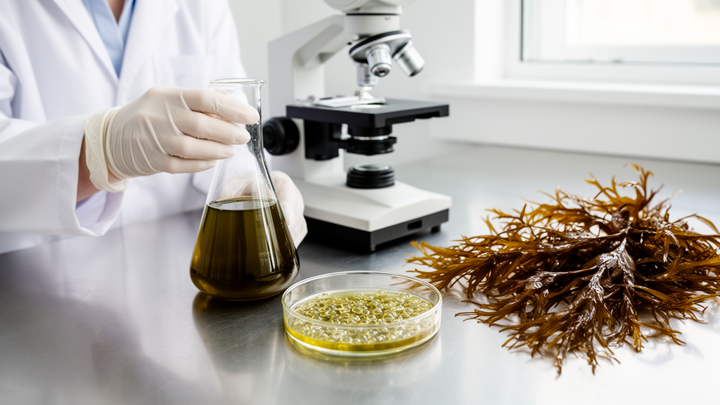
A brown seaweed that grows in dense, ribbon-like fronds along the Pacific coast of North America may hold the key to a new generation of anti-inflammatory therapies. In a study published in Molecular Biology Reports, researchers from Universidad Veracruzana and Universidad Autónoma de Baja California report that extracts of Egregia menziesii, commonly known as feather boa kelp, can calm overactive immune cells in the laboratory, dialing down two of the most important molecular drivers of inflammation. The work is the first systematic investigation of how this seaweed affects the inflammatory machinery of macrophages, the white blood cells that act as both first responders and long-term regulators of the immune system.
The research team, led by Omar Arroyo-Xochihua, focused on RAW 264.7 macrophages, a murine cell line that has become the workhorse of inflammation research worldwide. These cells were stimulated with lipopolysaccharide, or LPS, a molecule found in the outer membrane of Gram-negative bacteria that reliably triggers a full-blown inflammatory response. Once the macrophages were activated in this way, the scientists treated them with an ethanol-water extract of E. menziesii and with four fractions separated by polarity: hexane, dichloromethane, ethyl acetate, and a residual aqueous fraction. The goal was to see whether any of these preparations could rein in the cellular storm that LPS ignites.
Before any biological testing could begin, the team needed to know what was actually inside the seaweed. Using proton nuclear magnetic resonance spectroscopy, or 1H-NMR, they generated chemical fingerprints of the crude extract and each fraction. The spectra revealed a rich assortment of proton signals spanning alkyl, hydroxyl, amino, alkene, and aromatic regions. Three regions stood out. Signals between 1.10 and 1.25 parts per million corresponded to the methyl groups of fucoidan, a sulfated polysaccharide abundant in brown seaweed cell walls. Signals between 5.0 and 5.8 parts per million marked the anomeric protons of fucoidan’s sugar rings. And a dense cluster between 3 and 5 parts per million pointed to mannitol, a sugar alcohol that brown algae use as a carbon transport molecule and osmolyte.
Safety came first. Using the MTT assay, which measures mitochondrial activity as a proxy for cell health, the researchers exposed macrophages to concentrations ranging from 12.5 to 1000 micrograms per milliliter over 24 and 48 hours. The extract and all fractions kept cell viability at or above 90 percent across the tested range, and the calculated half-maximal inhibitory concentration exceeded 1000 micrograms per milliliter at both time points. Only the highest concentration of the crude extract showed any hint of toxicity at 48 hours, and it was excluded from further experiments. Hydrogen peroxide, used as a positive cytotoxicity control, killed cells as expected, confirming the assay was working. The result establishes that the seaweed preparations are safe for cellular experimentation at the doses that produced biological effects.
With safety established, the team turned to a scratch assay, a simple but informative test of cell migration. A sterile pipette tip drags a wound-like gap through a confluent monolayer of macrophages, and researchers then measure how quickly cells migrate in to close the gap. LPS alone impaired migration, leaving the scratched area largely open at 48 hours. Treatment with the crude ethanol-water extract significantly reversed this effect, reducing the free scratched area by up to 40 percent at 24 hours compared with the LPS-stimulated control. The aqueous fraction performed even better, showing significant scratch closure at both 24 and 48 hours, with reductions approaching 10 percent of the original wound area remaining at the later time point. Curcumin and indomethacin, included as natural and pharmacological controls, also promoted closure, but the seaweed treatments held their own against these benchmarks.
The most technically ambitious part of the study came next. Rather than guessing which compounds were responsible, the researchers applied multivariate data analysis, specifically Orthogonal Partial Least-Squares modeling, to correlate the NMR spectra with the scratch closure measurements. The resulting statistical model was strikingly robust, with explained variance values above 0.95 and predictive Q-squared values of 0.97 and 0.93 at 24 and 48 hours respectively, and cross-validation confirmed the model was not overfitted. The contribution plots revealed that proton signals in the 0.6 to 3 parts per million region were the main contributors to the anti-migratory rescue effect. This region encompasses the methyl groups of fucoidan and the alcohol protons of mannitol, providing chemometric evidence that these two abundant seaweed metabolites are the likely active principles.
Phagocytosis, the process by which macrophages engulf debris and pathogens, told a more nuanced story. Measured with a neutral red uptake assay, phagocytic activity surged in LPS-stimulated cells, as expected. The crude extract brought this activity back down below the levels seen in unstimulated controls, suggesting a normalizing rather than suppressive effect. Interestingly, the dichloromethane, ethyl acetate, and aqueous fractions each increased phagocytic activity above unstimulated baseline levels, hinting that different components of the seaweed may push macrophage behavior in different directions depending on the chemical context. The hexane fraction showed no significant effect either way.
The clearest anti-inflammatory signal emerged from the cytokine measurements. Using reverse transcription quantitative PCR and enzyme-linked immunosorbent assays, the team quantified both messenger RNA and secreted protein levels of interleukin-1 beta and interleukin-6, two pro-inflammatory cytokines central to the M1 macrophage phenotype that LPS induces. The crude extract cut IL-1 beta mRNA expression to less than half of the LPS-stimulated level and kept IL-6 fold change below 3, with corresponding reductions at the protein level that matched the effect of indomethacin. The aqueous fraction also significantly reduced both cytokines, though its protein-level effects were less complete. Notably, neither curcumin nor indomethacin achieved significant reductions in cytokine gene expression in this system, making the seaweed’s performance at the transcript level particularly noteworthy.
The mechanistic story fits with a growing body of literature on sulfated polysaccharides. Fucoidan’s negatively charged sulfate groups are thought to interact electrostatically with inflammatory mediators, and studies of fucoidan from related brown seaweeds have shown that it can block the NF-kappa B signaling pathway, a master transcriptional switch for inflammatory genes. Mannitol, meanwhile, has been shown to reduce markers of M1-like macrophage and microglial activation in animal models of neuroinflammation, possibly through osmotic effects that alter immune cell behavior. The authors caution that the compounds in a crude extract may act synergistically or even antagonistically, and that a single murine cell line in a monolayer cannot capture the full complexity of inflammation in a living organism. Future work, they suggest, should test human cell lines, explore nitric oxide and other inflammatory mediators, and pursue full metabolomic profiling.
Still, the implications are considerable. Non-steroidal anti-inflammatory drugs remain among the most widely consumed medications on Earth, and their long-term use is linked to gastrointestinal damage, ulcers, and elevated cardiovascular risk. Marine organisms remain a strikingly underexplored source of alternatives; between 1981 and 2019, more than fifty natural products and derivatives for treating inflammation were approved, yet marine species account for only a small fraction of the research effort. Egregia menziesii, with its limited range from Alaska to Baja California and its documented safety across multiple cell types and whole-organism models, now joins the shortlist of seaweeds with credible immunomodulatory credentials. If the fucoidan and mannitol hypothesis holds up in more complex systems, the feather boa kelp swaying in the Pacific surf could one day contribute to medicines that quiet inflammation without the collateral damage of current drugs.
Subject of Research: Anti-inflammatory activity and chemical profiling of Egregia menziesii extracts in LPS-activated macrophages
Article Title: Chemical profiling of Egregia menziesii and anti-inflammatory potential in LPS-activated RAW 264.7 macrophages
Article References: Arroyo-Xochihua, O., Sánchez-Medina, A., López-Franco, Ó., Olivares-Bañuelos, T. N., & Zepeda, R. C. (2026). Chemical profiling of Egregia menziesii and anti-inflammatory potential in LPS-activated RAW 264.7 macrophages. Molecular Biology Reports, 53(1), Article 1529. https://doi.org/10.1007/s11033-026-12685-y
Image Credits: AI Generated
DOI: 10.1007/s11033-026-12685-y
Keywords: Egregia menziesii, brown seaweed, fucoidan, mannitol, macrophages, inflammation, cytokines, 1H-NMR, chemometrics, RAW 264.7, sulfated polysaccharides, natural products
News Source: Drew Townsend. (October 19, 2026). Feather Boa Kelp Extract Shows Anti-Inflammatory Power in Lab-Grown Immune Cells. Scienmag.