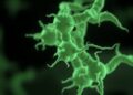
Averting our eyes from things that scare us may be due to a specific cluster of neurons in a visual region of the brain, according to new research at the University of Tokyo. Researchers found that in fruit fly brains, these neurons release a chemical called tachykinin which appears to control the fly’s movement to avoid facing a potential threat. Fruit fly brains can offer a useful analogy for larger mammals, so this research may help us better understand our own human reactions to scary situations and phobias. Next, the team want to find out how these neurons fit into the wider circuitry of the brain so they can ultimately map out how fear controls vision.
Credit: 2023, Tsuji et al.
Averting our eyes from things that scare us may be due to a specific cluster of neurons in a visual region of the brain, according to new research at the University of Tokyo. Researchers found that in fruit fly brains, these neurons release a chemical called tachykinin which appears to control the fly’s movement to avoid facing a potential threat. Fruit fly brains can offer a useful analogy for larger mammals, so this research may help us better understand our own human reactions to scary situations and phobias. Next, the team want to find out how these neurons fit into the wider circuitry of the brain so they can ultimately map out how fear controls vision.
Do you cover your eyes during horror movies? Or perhaps the sight of a spider makes you turn and run? Avoiding looking at things which scare us is a common experience, for humans and animals. But what actually makes us avert our gaze from the things we fear? Researchers have found that it may be due to a group of neurons in the brain which regulates vision when feeling afraid.
“We discovered a neuronal mechanism by which fear regulates visual aversion in the brains of Drosophila (fruit flies). It appears that a single cluster of 20-30 neurons regulates vision when in a state of fear. Since fear affects vision across animal species, including humans, the mechanism we found may be active in humans as well,” explained Assistant Professor Masato Tsuji from the Department of Biological Sciences at the University of Tokyo.
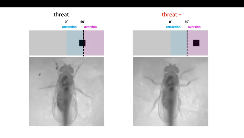
The team used puffs of air to simulate a physical threat and found that the flies’ walking speed increased after being puffed at. The flies also would choose a puff-free route if offered, showing that they perceived the puffs as a threat (or at least preferred to avoid them). Next the researchers placed a small black object, roughly the size of a spider, 60 degrees to the right or left of the fly. On its own the object didn’t cause a change in behavior, but when placed following puffs of air, the flies avoided looking at the object and moved so that it was positioned behind them.
To understand the molecular mechanism underlying this aversion behavior, the team then used mutated flies in which they altered the activity of certain neurons. While the mutated flies kept their visual and motor functions, and would still avoid the air puffs, they did not respond in the same fearful manner to visually avoid the object.
“This suggested that the cluster of neurons which releases the chemical tachykinin was necessary for activating visual aversion,” said Tsuji. “When monitoring the flies’ neuronal activity, we were surprised to find that it occurred through an oscillatory pattern, i.e., the activity went up and down similar to a wave. Neurons typically function by just increasing their activity levels, and reports of oscillating activity are particularly rare in fruit flies because up until recently the technology to detect this at such a small and fast scale didn’t exist.”
By giving the flies genetically encoded calcium indicators, the researchers could make the flies’ neurons shine brightly when activated. Thanks to the latest imaging techniques, they then saw the changing, wavelike pattern of light being emitted, which was previously averaged out and missed.
Next, the team wants to figure out how these neurons fit into the broader circuitry of the brain. Although the neurons exist in a known visual region of the brain, the researchers do not yet know from where the neurons are receiving inputs and to where they are transmitting them, to regulate visual escape from objects perceived as dangerous.
“Our next goal is to uncover how visual information is transmitted within the brain, so that we can ultimately draw a complete circuit diagram of how fear regulates vision,” said Tsuji. “One day, our discovery might perhaps provide a clue to help with the treatment of psychiatric disorders stemming from exaggerated fear, such as anxiety disorders and phobias.”
#####
Paper Title:
Masato Tsuji, Yuto Nishizuka, Kazuo Emoto. Threat gates visual aversion via theta activity in Tachykinergic neurons. Nature Communications. doi: 10.1038/s41467-023-39667-z
Funding
This research was supported by the Japan Society for the Promotion of Science (JSPS) through the Graduate Program for Leaders in Life Innovation (GPLLI), MEXT Grants-in-Aid for Scientific Research on Innovative Areas “Dynamic regulation of brain function by Scrap and Build system” (KAKENHI 16H06456), JSPS (KAKENHI 16H02504), WPI-IRCN, AMED-CREST (JP21gm1310010), JST-CREST (JPMJCR22P6), Toray Foundation, Naito Foundation, Takeda Science Foundation, and Uehara Memorial Foundation.
Useful Links
Graduate School of Science: https://www.s.u-tokyo.ac.jp/en/
International Research Center for Neurointelligence: https://ircn.jp/en/
Research Contacts:
Assistant Professor Masato Tsuji
Department of Biological Sciences
Graduate School of Science
The University of Tokyo
7-3-1 Hongo, Bunkyo-ku, Tokyo 113-0033, Japan
Email: [email protected]
Tel.:+81-3-5841-4427
Professor Kazuo Emoto
Department of Biological Sciences
Graduate School of Science
The University of Tokyo
7-3-1 Hongo, Bunkyo-ku, Tokyo 113-0033, Japan
Email: [email protected]
Tel.: +81-3-5841-4426
Press contact:
Mrs. Nicola Burghall
Public Relations Group, The University of Tokyo,
7-3-1 Hongo, Bunkyo-ku, Tokyo 113-8654, Japan
[email protected]
About the University of Tokyo
The University of Tokyo is Japan’s leading university and one of the world’s top research universities. The vast research output of some 6,000 researchers is published in the world’s top journals across the arts and sciences. Our vibrant student body of around 15,000 undergraduate and 15,000 graduate students includes over 4,000 international students. Find out more at www.u-tokyo.ac.jp/en/ or follow us on Twitter at @UTokyo_News_en.
Journal
Nature Communications
DOI
10.1038/s41467-023-39667-z
Method of Research
Experimental study
Subject of Research
Animals
Article Title
Threat gates visual aversion via theta activity in Tachykinergic neurons.
Article Publication Date
13-Jul-2023