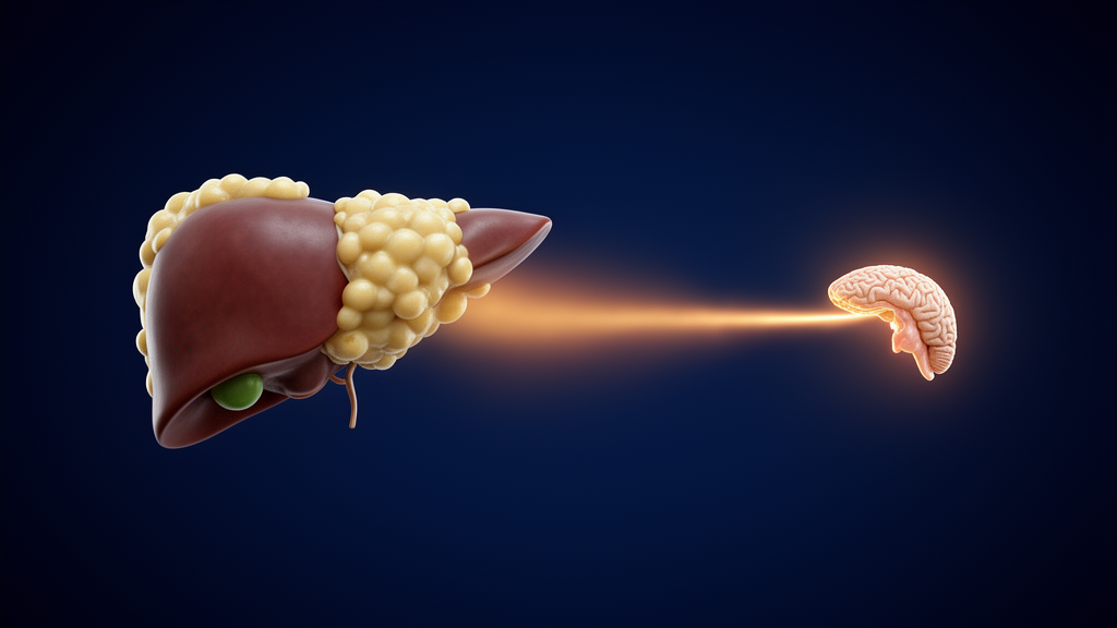
People with metabolic dysfunction-associated steatotic liver disease (MASLD) show a flattened overnight rise in thyrotropin, the pituitary hormone better known as TSH, according to a prospective cross-sectional study published in BMC Endocrine Disorders. In a single-centre investigation of hospitalised adults, researchers led by Yasemin Çöl measured thyroid function at 08:00 and again at 24:00 in 53 patients with ultrasound-defined steatosis fulfilling MASLD criteria and in 32 controls without steatosis. While TSH in the control group climbed noticeably over the night, patients with MASLD showed essentially no such rise, a difference the authors describe as hypothesis-generating and in need of confirmation in ambulatory cohorts.
The biological rationale for the measurement is grounded in liver physiology. The liver carries out most peripheral thyroid hormone metabolism, and TSH secretion normally rises overnight. This nocturnal surge is known to be blunted in non-thyroidal illness, but it had not previously been examined in MASLD. The primary analysis was a linear mixed model of TSH with a group-by-time interaction, and the summary measure was the midnight-minus-morning difference in TSH, abbreviated ΔTSH.
The results showed a statistically detectable difference between the groups. The interaction term was −0.49 mU/L (95% confidence interval −0.96 to −0.01; p = 0.044), indicating that the overnight trajectory of TSH diverged between MASLD patients and controls. Median ΔTSH was −0.14 mU/L in the MASLD group versus 0.19 mU/L in controls (p = 0.019), meaning TSH failed to rise significantly overnight in the patients. Notably, the attenuation reflected mainly a higher morning TSH in the MASLD group (1.40 vs. 1.18 mU/L; p = 0.020) rather than a lower midnight value, so the flattened curve was anchored at its morning end.
Interpretation is complicated by the metabolic profile of the patients, who had higher body mass index, higher glycated haemoglobin and a far greater prevalence of diabetes than the controls (66.0% vs. 21.9%). When the authors adjusted the regression for age, sex, BMI and diabetes, the difference remained similar in size but lost statistical significance (β −0.52, 95% CI −1.09 to 0.06; p = 0.076), and its independence from diabetes could not be established. After further adjustment for anti-thyroid peroxidase antibodies, the estimate was again significant (β −0.59, 95% CI −1.11 to −0.06).
Stratified analyses sharpened the picture further. ΔTSH was close to zero in diabetic participants of both groups, and among participants without diabetes it was lower in the MASLD group, although the non-parametric comparison did not reach significance (t test p = 0.028; Mann–Whitney p = 0.097). In a post hoc analysis, the group difference appeared only when C-reactive protein exceeded 10 mg/L (interaction p = 0.050), suggesting that systemic inflammation may coexist with the attenuated rise.
Two further findings frame the clinical meaning of the result. The ΔTSH measure discriminated MASLD poorly, with an area under the curve of 0.653, so the authors do not present it as a diagnostic tool. Separately, midnight free triiodothyronine correlated inversely with the NAFLD fibrosis score (r = −0.54; q < 0.001), an association that hints at a link between lower nighttime free T3 and fibrosis-related risk scoring, though the correlation alone cannot establish direction or mechanism.
The study design carries limitations that the authors acknowledge directly. It was single-centre and restricted to hospitalised adults, a population in which acute illness itself can alter thyroid hormone dynamics, and the cross-sectional design cannot establish causal relationships between liver steatosis, metabolic dysfunction and TSH regulation. The attenuation of the nocturnal TSH rise coexisted with dysglycaemia and, in the post hoc analysis, with systemic inflammation, but the study could not determine whether these factors explain the finding or merely accompany it.
The authors conclude that in hospitalised patients with MASLD the nocturnal TSH rise was attenuated, and that these hypothesis-generating findings require confirmation in ambulatory cohorts before any clinical application can be considered. Until such confirmation is available, the result stands as an observational signal linking hepatic steatosis to the circadian regulation of the pituitary-thyroid axis rather than as a basis for testing or treatment decisions.
Subject of Research: Attenuated nocturnal thyrotropin rise in metabolic dysfunction-associated steatotic liver disease
Article Title: Attenuated nocturnal thyrotropin rise in metabolic dysfunction-associated steatotic liver disease: a prospective cross-sectional study
Article References: Çöl, Y., Yakut Suner, M., Çöl, M., Erişmiş, B., & Öğmen, B. (2026). Attenuated nocturnal thyrotropin rise in metabolic dysfunction-associated steatotic liver disease: a prospective cross-sectional study. BMC Endocrine Disorders. https://doi.org/10.1186/s12902-026-02689-x
Image Credits: AI Generated
DOI: 10.1186/s12902-026-02689-x
Keywords: MASLD, thyrotropin, TSH, hepatic steatosis, circadian rhythm, thyroid hormone metabolism, diabetes, C-reactive protein, cross-sectional study, NAFLD fibrosis score, endocrinology, Attenuated
News Source: Ophelia Keating. (October 26, 2026). Fatty liver disease blunts the overnight rise of thyroid-stimulating hormone. Scienmag.