


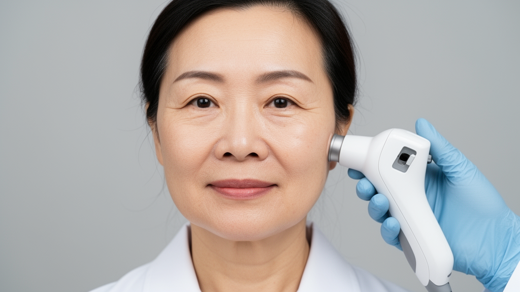
A cross-sectional study of 547 Chinese women aged 18 to 65 years has mapped how the microscopic architecture of facial skin changes across adulthood, finding that measurable remodeling of the dermis and the dermo-epidermal junction becomes prominent from the 36-to-45-year age group onward. The research, published in Dermatology and Therapy, combined two-photon microscopy with high-frequency ultrasound to capture structural parameters alongside biophysical measurements and clinical aging phenotypes across four facial zones.
Participants were recruited between March and May 2024 from three cities—Shanghai, Guangzhou, and Luoyang—using a stratified quota sampling method across five predefined age brackets: 18–25, 26–35, 36–45, 46–55, and 56–65 years. The study applied strict inclusion and exclusion criteria to reduce confounding. Participants were required to have an average daily sleep duration of at least six hours over the previous six months, daily sun exposure of no more than four hours, and consistent sun protection habits. Those who had undergone dermatologic or aesthetic procedures, such as energy-based devices, injectables, or oral collagen, within a six-month washout period were excluded, as were pregnant or lactating women, people with severe systemic or endocrine disorders, oral contraceptive users, and those using topical or oral nutritional or cosmetic products known to affect skin outcomes within two weeks prior to the study.
During a single study visit, researchers measured structural, biophysical, and clinical skin parameters on the forehead, glabella, eye corner, and cheek. Two-photon microscopy, operating at an excitation wavelength of 780 nanometers with laser power kept at or below 500 milliwatts to avoid photodamage, captured second harmonic generation (SHG) and autofluorescence (AF) signals. SHG reflects the density of collagen, while AF represents the abundance of elastic fibers, or elastotic material. From these signals the team derived the SHG-to-AF aging index of dermis (SAAID), the elastin-to-collagen ratio (ELCOR), and the dermo-epidermal junction index (DEJI), which captures the tortuosity and undulation of the junction that anchors the epidermis to the dermis. High-frequency ultrasound at 20 megahertz provided dermal density, dermal thickness, and echogenic pixel measures, with dermal density quantified as mean grayscale intensity within a manually delineated region of interest bounded by the epidermal entrance echo above and the dermo-hypodermal junction below.
The key age-associated structural differences included decreased SAAID and dermal density, increased elastin-to-collagen ratio, and flattening of the dermo-epidermal junction becoming prominent in the 36–45-year age group and beyond. SAAID showed a consistent decrease beginning with the 36–45-year group across facial zones, reflecting a decline in collagen relative to elastin in the dermal layer. ELCOR generally increased with age, although values dipped slightly between the 18–25 and 26–35-year groups before rising significantly from the 36–45-year group onward in the forehead and glabella. Dermal density declined in all facial zones, particularly from the 36–45-year group onward, with further reduction in older groups. The ratio of superior to inferior lower echogenic pixels increased from the 36–45-year group onward across all zones, indicating declining extracellular matrix density and integrity, while middle and high echogenic pixels decreased with age in the same period.
Zonal differences were pronounced. The forehead displayed the lowest SAAID values, followed by the glabella, both corresponding to the highest ELCOR values and therefore the lowest collagen-to-elastin ratios. The cheek showed the greatest variation across age groups among all facial zones, serving as what the authors describe as a clear example of the microstructural changes that occur with age. The eye corner exhibited the highest dermal density values, whereas the glabella displayed the lowest density across all age groups. Stratum corneum thickness generally increased with age while epidermal thickness decreased, with the forehead showing the highest and the eye corner the lowest values for both. Dermal thickness varied by zone rather than age, with cheeks showing the highest values. The glabella consistently exhibited the highest LEPs/LEPi ratio and the lowest MEP and HEP values, which the authors interpret as indicating reduced protein synthesis capacity and higher susceptibility to structural degradation.
Biophysical measurements told a parallel story. Skin elasticity, measured with a Cutometer, was progressively lower across older age groups, with the sharpest decline observed in the eye corner. Hydration and transepidermal water loss decreased with age across facial zones, particularly from the 36–45-year group onward. Skin lightness declined in all facial zones, while redness and yellowness increased, particularly in ultraviolet-exposed zones such as the cheeks and forehead. Phenotypical aging signs moved in the opposite direction: wrinkle severity and pigmentation grading increased with age. Crow’s feet wrinkle grading showed a very strong positive correlation with age (r = 0.916), the strongest among the clinical parameters in this study population, followed by glabella wrinkles (r = 0.862) and forehead wrinkles (r = 0.793). Pigmentation spot density at the cheek showed a moderate correlation (r = 0.582), as did the number of identified pigmentary spots (r = 0.662). All clinical assessments were performed by a single trained evaluator, blinded to participants’ ages, under consistent lighting conditions using a standardized atlas adapted from L’Oréal’s Skin Aging Atlas Volume 2: Asian Type.
To explore how structural parameters relate to surface-level characteristics, the researchers applied principal component analysis to the structural parameters of the cheek and eye corner zones. Two components emerged under the Kaiser criterion. Principal component 1 (PC1) was primarily influenced by positive loadings from dermal density, middle echogenic pixels, and high echogenic pixels, with the LEPs/LEPi ratio loading negatively, suggesting that this dimension reflects dermal echogenicity and extracellular matrix integrity. Principal component 2 (PC2) reflected the elastin-to-collagen profile, characterized by positive loadings from autofluorescence and negative loadings from SAAID. Together, PC1 and PC2 explained approximately 46 percent of the total variance in the cheek zone and 48 percent in the eye corner zone. Logistic regression on the principal components showed a positive association of PC1 with favorable skin characteristics such as elasticity, hydration, and lightness, and lower severity of wrinkles and hyperpigmentation (p < 0.05), while the inverse held for PC2 (p < 0.05). Higher PC2 scores were generally linked to less favorable characteristics, including reduced hydration, darker skin tone, increased pigmentation, decreased elasticity, and increased wrinkle severity.
The authors frame the findings as adding a microstructural dimension to previously reported nonlinear facial aging trajectories in Chinese populations, in which large-scale phenotypic analyses have identified the early 30s as a first phase of rapid aging followed by a second acceleration in the late 50s. Because those earlier studies relied primarily on phenotypic or biophysical measures, the authors suggest that microstructural deterioration may emerge slightly later than initial phenotypic divergence, with measurable remodeling of dermal structure becoming evident after the mid-30s. They caution that this temporal offset reflects differences in endpoint sensitivity rather than a conflict with prior literature, and they note that because participants were stratified into predefined age brackets, the exact timing of biological acceleration points could not be mathematically isolated; future work should analyze age as a continuous variable using segmented regression, spline regression, or formal change-point analysis.
Several other limitations deserve emphasis. The cross-sectional design captures differences between age groups at a single time point rather than changes within individuals over time. Participants came from three Chinese cities, and environmental factors may vary across regions, while data collection was limited to a single season to minimize seasonal variation. Intraobserver and interobserver reproducibility were not assessed because a single operator conducted the testing. The two-photon microscopy system imaged to a depth of 140 micrometers over a 140 by 140 micrometer field of view, precluding characterization of deeper dermal architecture and providing only localized sampling. The PCA model explained less than half of the total variance, indicating that additional structural dimensions not captured here also contribute to skin aging, and the authors call for further validation with a broader panel of dermal parameters. Within those constraints, the study offers a noninvasive structural reference for interpreting age-related collagen deterioration in Chinese women and supports the combined use of two-photon microscopy and ultrasound in dermatological aging assessment, with the authors noting that longitudinal studies will be required to determine whether layer-targeted interventions yield better outcomes.
Subject of Research: Age-associated differences in facial skin structure, biophysical properties, and visible aging signs in Chinese women assessed with two-photon microscopy and high-frequency ultrasound
Article Title: Age-Associated Differences in Facial Skin Structure in Chinese Women: a Cross-Sectional Study
Article References: Zhang, C., Wang, K., Gao, L., Wu, J., Melnikov, V., Wang, Y., Wang, H., Song, W., Wang, H., Guo, Z., Wang, X., Flament, F., Yang, B., Li, Q., Liu, F., & Xiang, L. (2026). Age-Associated Differences in Facial Skin Structure in Chinese Women: a Cross-Sectional Study. Dermatology and Therapy, 16(11), 5743-5759. https://doi.org/10.1007/s13555-026-01931-7
Image Credits: AI Generated
DOI: 10.1007/s13555-026-01931-7
Keywords: skin aging, two-photon microscopy, high-frequency ultrasound, dermis, collagen, elastin, dermo-epidermal junction, Chinese women, cross-sectional study, facial zones, dermal density, principal component analysis
News Source: Beatrice Stafford. (October 30, 2026). Facial Skin Aging in Chinese Women Shows Structural Shifts After the Mid-Thirties. Scienmag.



