Cell division is one of the most fundamental processes of life. From bacteria to blue whales, every living being on Earth relies on cell division for growth, reproduction, and species survival. Yet, there is remarkable diversity in the way different organisms carry out this universal process. A new study from EMBL Heidelberg’s Dey group and their collaborators, recently published in Nature, explores how different modes of cell division evolved in close relatives of fungi and animals, demonstrating, for the first time, the link between an organism’s life cycle and the way their cells divide.
Credit: ©Nirupa Rao
Cell division is one of the most fundamental processes of life. From bacteria to blue whales, every living being on Earth relies on cell division for growth, reproduction, and species survival. Yet, there is remarkable diversity in the way different organisms carry out this universal process. A new study from EMBL Heidelberg’s Dey group and their collaborators, recently published in Nature, explores how different modes of cell division evolved in close relatives of fungi and animals, demonstrating, for the first time, the link between an organism’s life cycle and the way their cells divide.
Despite last sharing a common ancestor over a billion years ago, animals and fungi are similar in many ways. Both belong to a broader group called ‘eukaryotes’ – organisms whose cells store their genetic material inside a closed compartment called the ‘nucleus’. The two differ, however, in how they carry out many physiological processes, including the most common type of cell division – mitosis.
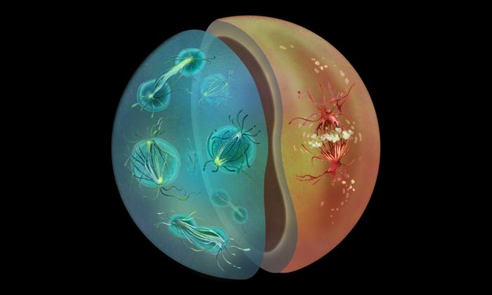
Most animal cells undergo ‘open’ mitosis, in which the nuclear envelope – the two-layered membrane separating the nucleus from the rest of the cell – breaks down when cell division begins. However, most fungi use a different form of cell division – called ‘closed’ mitosis – in which the nuclear envelope remains intact throughout the division process. However, very little is known about why or how these two distinct modes of cell division evolved and what factors determine which mode would be predominantly followed by a particular species.
This question captured the attention of scientists in the Dey Group at EMBL Heidelberg, who investigate the evolutionary origins of the nucleus and cell division. “By studying diversity across organisms and reconstructing how things evolved, we can begin to ask if there are universal rules that underlie how such fundamental biological processes work,” said Gautam Dey, Group Leader at EMBL Heidelberg.
In 2020, during the COVID-19 lockdown, an unexpected path to answering this question grew out of discussions between Dey’s group and Omaya Dudin’s team at the Swiss Federal Institute of Technology (EPFL), Lausanne. Dudin is an expert in an unusual group of marine protists – Ichthyosporea. Ichthyosporea are closely related to both fungi and animals, with different species lying closer to one or the other group on the evolutionary family tree.
The Dey and Dudin groups, in collaboration with Yannick Schwab’s group at EMBL Heidelberg, decided to probe the origins of open and closed mitosis using Ichthyosporea as a model. Interestingly, the researchers found that certain species of Ichthyosporea undergo closed mitosis while others undergo open mitosis. Therefore, by comparing and contrasting their biology, they could obtain insights into how organisms adapt to and use these two cell division modes.
Hiral Shah, an EIPOD fellow working across the three groups, led the study. “Having recognised very early that Ichthyosporea, with their many nuclei and key evolutionary position between animal and fungi, were well-suited for addressing this question, it was clear that this would require bringing together the cell biological and technical expertise of the Dey, Dudin, and Schwab groups, and this is exactly what the EIPOD fellowship allowed me to do,” said Shah.
Upon closely probing the mechanisms of cell division in two species of Ichthyosporeans, the researchers found that one species, S. arctica, favours closed mitosis, similar to fungi. S. arctica also has a life cycle with a multinucleate stage, where many nuclei exist within the same cell – another feature shared with many fungal species as well as the embryonic stages of certain animals, such as fruit flies. Another species, C. perkinsii, turned out to be much more animal-like, relying on open mitosis. Its life cycle involves primarily mononucleate stages, where each cell has a single nucleus.
“Our findings led to the key inference that the way animal cells do mitosis evolved hundreds of millions of years before animals did. The work therefore has direct implications for our general understanding of how eukaryotic cell division mechanisms evolve and diversify in the context of diverse life cycles, and provides a key piece of the animal origins puzzle,” said Dey.
The study combined expertise in comparative phylogenetics, electron microscopy (from the Schwab Group and the electron microscopy core facility (EMCF) at EMBL Heidelberg), and ultrastructure expansion microscopy, a technique that involves embedding biological samples in a transparent gel and physically expanding it. Additionally, Eelco Tromer, from the University of Groningen in the Netherlands, and Iva Tolic, from the Ruđer Bošković Institute in Zagreb, Croatia, provided expertise in comparative genomics and mitotic spindle geometry and biophysics, respectively.
“The first time we saw an expanded S. arctica nucleus, we knew this technique would change the way we study the cell biology of non-model organisms,” said Shah, who brought back the expansion microscopy technique to EMBL Heidelberg after a stint at the Dudin lab. Dey agrees: “A key breakthrough in this study came with our application of ultrastructure expansion microscopy (U-ExM) to the analysis of the ichthyosporean cytoskeleton. Without U-ExM, immunofluorescence and most dye labelling protocols do not work in this understudied group of marine holozoans.”
This study also demonstrates the importance of going beyond traditional model organism research when trying to answer broad biological questions, and the potential insights further research on Ichthyosporean systems might reveal. “Ichthyosporean development displays remarkable diversity,” said Dudin. “On one hand, several species exhibit developmental patterns similar to those of early insect embryos, featuring multinucleated stages and synchronised cellularisation. On the other hand, C. perkinsii undergoes cleavage division, symmetry breaking, and forms multicellular colonies with distinct cell types, similar to the ‘canonical view’ of early animal embryos. This diversity not only helps in understanding the path to animals but also offers a fascinating opportunity for comparative embryology outside of animals, which is, in itself, very exciting.”
The project’s inherent interdisciplinarity served not only as a good testbed for this type of collaborative research but also for the unique postdoctoral training afforded at EMBL. “Hiral’s project nicely illustrates the virtue of the EIPOD programme: a truly interdisciplinary project, bundling innovative biology with advanced methods, all contributing to a truly spectacular personal development,” said Schwab. “We (as mentors) witnessed the birth of a strong scientist, and this is really rewarding!”
The Dey, Dudin, and Schwab groups are currently also collaborating on the PlanExM project, part of the TREC expedition – an EMBL-led initiative to explore and sample the biodiversity along European coasts. PlanExM aims to apply expansion microscopy to study the ultrastructural diversity of marine protists directly in environmental samples. “The project grew out of the realisation that U-ExM is going to be a game-changer for protistology and marine microbiology,” said Dey. With this project, as well as others currently underway, the research team hopes to shed further light on the diversity of life on Earth and the evolution of the fundamental biological processes.
Journal
Nature
DOI
10.1038/s41586-024-07430-z
Subject of Research
Animals
Article Title
Life cycle-coupled evolution of mitosis in close relatives of animals
Article Publication Date
22-May-2024
