
Predictive imaging technology in the realm of acute ischemic stroke has taken significant strides forward, especially regarding the management of patients experiencing large vessel obstruction (AIS-LVO). A study published in BioMedical Engineering OnLine shed light on the relationship between CT imaging features and the risk of hemorrhagic transformation (HT) following mechanical thrombectomy in patients suffering from AIS-LVO. This pioneering research aims to establish a connection between pre-existing medical conditions, imaging characteristics, and the risk of complications post-thrombectomy, potentially transforming clinical practices and patient outcomes.
The research embarked on a critical investigation into whether specific CT imaging features could serve as reliable predictors for hemorrhagic transformation in patients undergoing thrombectomy. The primary goal was to evaluate various clinical and CT imaging factors that might indicate a heightened risk of HT, particularly in the first 24 hours following the procedure. By discerning these predictive factors, the study addresses an urgent need for enhanced monitoring and preventative measures for vulnerable patient populations facing acute ischemic events.
In conducting this study, a cohort of 135 patients diagnosed with AIS-LVO at a single hospital facility between August 2021 and May 2023 was retrospectively analyzed. All the subjects underwent the standardized mechanical thrombectomy procedure, following which the patients were stratified into two distinct groups: those who experienced hemorrhagic transformation (HT group) and those who did not (non-HT group). This rigorous classification allowed for a focused exploration of the variables affecting post-operative outcomes and recovery trajectories for both cohorts.
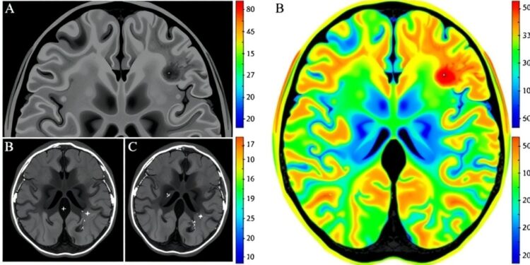
Following the intervention, all participants in both groups underwent a comprehensive CT examination. The objective was to meticulously document and assess the changes in CT imaging indicators between the HT and non-HT groups. Key variables evaluated included the history of atrial fibrillation, the NIH Stroke Scale (NIHSS) scores, galectin-3 (Gal-3) levels, and various CT imaging features such as time to peak (TTP), cerebral blood flow (CBF), and specific signs like the Hyperdense Middle Cerebral Artery Sign (HMCAS). These factors collectively form a framework through which researchers dissect and comprehend the multifaceted implications of CT imaging in acute stroke management.
Statistical analyses, particularly logistic regression, were employed to elucidate the correlations and impact of these imaging and clinical factors on the occurrence of HT. The results unveiled a striking association between the history of atrial fibrillation and heightened risk of hemorrhagic transformation. Furthermore, significant increases in NIHSS scores and Gal-3 levels were documented in patients who experienced HT, which may suggest a need for more proactive and targeted medical interventions in those presenting with these clinical indicators.
In terms of CT imaging findings, the study reported that the HT group exhibited a significantly higher prevalence of contrast medium exudation and HMCAS, alongside an extended TTP and decreased CBF. These imaging characteristics not only reinforced existing clinical understandings of acute stroke but also provided new insights into how these parameters correlate with post-thrombectomy complications. The data emphasizes the potential for CT imaging to function as a predictive tool rather than merely a diagnostic one, integrating real-time physiological assessments into patient management protocols.
Crucially, the research team developed a logistic regression model to calculate the likelihood of HT following thrombectomy. The model synthesized multiple predictive factors, including atrial fibrillation history, NIHSS scores prior to thrombectomy, Gal-3 levels, and various CT imaging features. It demonstrated a remarkable predictive accuracy, with an area under the curve (AUC) of 0.873, signifying a high sensitivity and specificity for identifying patients at risk of HT. Such a model could prove invaluable for clinicians in making informed decisions regarding patient care post-thrombectomy.
The implications of these findings extend beyond academic curiosity. By integrating predictive CT imaging features into clinical strategy, it may be possible to refine patient selection for thrombectomy, enhance postoperative monitoring, and ultimately decrease the incidence of hemorrhagic transformations. This forward-thinking approach could pave the way for personalized medicine in acute ischemic stroke interventions, where treatments and care protocols are tailored to individual patient needs based on tangible predictive parameters.
Moreover, the findings suggest that isolated CT imaging features such as TTP and CBF possess their diagnostic abilities, with AUC values recorded as 0.728 and 0.736, respectively. However, the combined assessment of these parameters demonstrated superior diagnostic capabilities, yielding an AUC of 0.783. Such synergy underscores the importance of integrated imaging assessments in clinical practice, particularly within the critical context of acute stroke management, where timely intervention can drastically alter patient outcomes.
In conclusion, this research not only illuminates the intricate interplay between CT imaging metrics and the heightened risks of hemorrhagic transformation but also proposes a forward-thinking model that could radically transform clinical approaches to stroke management. By focusing on predictive indicators that are readily identifiable in the clinical setting, healthcare providers can enhance the quality of care, optimize recovery, and refine tactical responses to potential complications in patients undergoing mechanical thrombectomy for AIS-LVO.
As the medical community continues to delve deeper into the importance of predictive analytics and imaging technologies, this study stands as a notable advancement. It highlights the transformative potential of leveraging CT findings to navigate the complexities of acute ischemic stroke treatment, ultimately aiming to reduce morbidity and mortality associated with hemorrhagic transformation in vulnerable populations.
Subject of Research: Predictive value of CT imaging features on the risk of hemorrhagic transformation after mechanical thrombectomy for acute ischemic stroke with large vessel obstruction
Article Title: Predictive value of CT imaging features on the risk of hemorrhagic transformation after mechanical thrombectomy for acute ischemic stroke with large vessel obstruction
News Publication Date: 2025
Web References:
References:
Image Credits:
Keywords: Acute ischemic stroke, CT imaging, Hemorrhagic transformation, Mechanical thrombectomy, Predictive model, Atrial fibrillation, Cerebral blood flow, Time to peak, Hyperdense Middle Cerebral Artery Sign, Galectin-3, NIHSS, Vascular obstruction.
Tags: acute ischemic stroke managementclinical factors in strokecomplications post-thrombectomyCT imaging characteristicshemorrhagic transformation riskimaging predictors of complicationslarge vessel occlusion predictorsmechanical thrombectomy outcomespatient monitoring strategiespredictive imaging technologyretrospective analysis of AIS-LVOstroke patient outcomes

