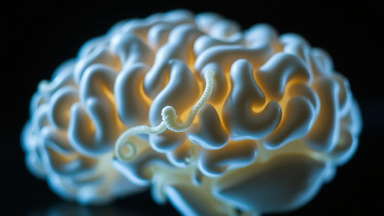
Human brain organoids have rapidly become one of the most compelling tools in modern neuroscience, offering researchers a three-dimensional, laboratory-grown glimpse into processes that were previously hidden inside the developing skull. A new review published in Bioengineering & Translational Medicine argues, however, that the field has reached a critical inflection point: the usefulness of a brain organoid can no longer be judged by how much it looks like brain tissue, but only by whether its engineering design matches the biological question being asked and whether that match is supported by rigorous functional evidence. The authors, led by researchers including Guohong Huang, Chenfei Lu, and senior investigators Guixue Wang and Chuanrong Zhao, propose an engineering-to-function framework that links every controllable construction variable, from cell source to culture platform, with the specific validation readouts required to make credible claims.
The review begins with the fundamentals. Brain organoids are generated from human pluripotent stem cells, either embryonic stem cells or induced pluripotent stem cells, which aggregate into embryoid bodies and self-organize into neural tissue. In unguided protocols, these aggregates rely on intrinsic developmental cues, producing heterogeneous tissues that can contain features of several brain regions, including cortical, telencephalic, and choroid plexus-like identities. Guided protocols, by contrast, apply defined morphogens that modulate key signaling pathways such as WNT, sonic hedgehog, FGF, BMP, and retinoic acid to steer cells toward cortical, ventral forebrain, midbrain, hypothalamic, or choroid plexus-like lineages. The resulting tissues can recapitulate striking features of early cortical development, including ventricular-like lumens lined by SOX2-, PAX6-, and NESTIN-positive neural progenitors, from which differentiating neurons migrate outward toward cortical plate-like zones that echo the layered architecture of the human cortex.
Yet morphology, the authors stress, is a deceptive benchmark. The construction variables that shape an organoid’s identity include the cell source and genetic background, extracellular matrix support, embryoid body formation method, patterning strategy, culture platform, vascularization approach, and the incorporation of additional cell types such as astrocytes, microglia, or endothelial cells. Each choice ripples through regional identity, cellular composition, maturation state, and viability. Matrigel, the most widely used matrix, supports neuroepithelial expansion and three-dimensional morphogenesis, but its undefined composition and batch-to-batch variability actively undermine reproducibility. The review calls for defined hydrogels with tunable stiffness, degradability, and ligand composition as more controllable alternatives for future organoid engineering, framing this substitution as a prerequisite for translational standardization rather than a cosmetic improvement.
Culture conditions compound these challenges. Because organoids lack vasculature, oxygen and nutrients must diffuse inward from the surrounding medium, and interior cells frequently succumb to hypoxia and necrosis during long-term culture. Suspension and rotating bioreactor systems, first popularized in the landmark 2013 protocols, improve medium mixing and mass transfer, but dish-based approaches still provide limited control over aggregate size and oxygen gradients, contributing to inter-organoid variability. Microfluidic organoid-on-a-chip platforms go further, using miniaturized channels, valves, and sensors to regulate flow, compartmentalization, temperature, pH, nutrient supply, and drug exposure with unprecedented precision. Pillar and perfusion plate systems offer a parallel, higher-throughput middle ground, with evidence that continuous flow can improve oxygen supply and reduce necrotic regions compared with static culture. The review cautions, however, that every engineering benefit must be demonstrated with quantitative readouts, including organoid size distribution, hypoxia markers, viability, lineage composition, and electrophysiological maturation, rather than assumed from device sophistication.
Nowhere is the gap between engineering ambition and functional evidence more pronounced than in vascularization. The living brain is an energy-hungry organ wrapped in a dense capillary network, and avascular organoids inevitably suffer restricted proliferation, premature differentiation, impaired neurogenesis, and aberrant cortical patterning. The review details two principal strategies for closing this gap. In vitro, researchers embed endothelial cells in the matrix surrounding organoids, induce the endothelial transcription factor ETV2 to generate vascular-like networks, or fuse brain organoids with vascular spheroids, with Wnt/β-catenin pathway activation further promoting vessel formation. In vivo, transplantation into rodent brains allows host vasculature to infiltrate the graft, with vascular ingrowth detectable within 7 to 10 days and robust blood flow confirmed by two-photon microscopy. In one striking study, transplanted human brain organoids survived for up to 233 days in mice, with 85.4 percent of grafts becoming vascularized, while nonvascularized organoids failed to survive; vascularized grafts showed reduced apoptosis, larger size, and greater numbers of mature neurons.
The authors draw a sharp line, however, between vascularization and functional blood-brain barrier formation. Endothelial-like cells inside organoids may express tight-junction proteins and transporter markers such as Claudin-5, GLUT1, and P-gp, but calling this a reconstructed BBB requires permeability assays, transendothelial electrical resistance measurements, and transporter activity studies. Barrier maturation in vivo depends on orchestrated signaling, including Wnt/β-catenin for endothelial specification, PDGF-B/PDGFRβ for pericyte recruitment, and sonic hedgehog for barrier integrity, and no current organoid platform reproduces the full cellular composition, perfusion dynamics, or regional heterogeneity of the human neurovascular unit. The review’s message is that strong claims about BBB-like function demand multi-level evidence, not marker expression alone.
Functional validation receives an equally systematic treatment. Morphological quality control should track size, volume, surface regularity, neuroepithelial bud formation, ventricular zone-like structures, and necrotic or cystic regions, but these endpoints cannot establish regional identity or synaptic function. For that, the review turns to electrophysiology. Patch-clamp recording resolves single-cell excitability, revealing, for example, functionally mature neurons with voltage-dependent sodium and potassium currents in microglia-containing organoids and pacemaking dopaminergic neurons in midbrain-like models relevant to Parkinson’s disease. Multielectrode arrays provide non-invasive, longitudinal recordings of spikes, local field potentials, network bursts, and synchrony, capturing the emergence of network-level activity during long-term culture, including epileptiform-like dynamics in organoids derived from patients with neurodevelopmental disorders. Imaging adds another layer: marker panels spanning SOX2 and Ki67 for progenitors, DCX and TUJ1 for immature neurons, MAP2 and NeuN for mature neurons, and GFAP and IBA1 for astrocytes and microglia, benchmarked increasingly against single-cell transcriptomic references of the developing human brain.
The translational payoff of this framework is illustrated across a remarkable range of applications. TP53 knockdown organoids revealed disorganized neural stem cell layers and cell-cycle accumulation in G1 phase, illuminating the tumor suppressor’s role in human brain development. CDK5RAP2-mutant organoids reproduced the microcephaly phenotype with reduced volume and thinner neuroepithelium. Patient-derived midbrain organoids carrying the LRRK2 G2019S Parkinson’s mutation showed impaired dopaminergic markers, phosphorylated α-synuclein accumulation, and elevated mitophagy, with an LRRK2 kinase inhibitor rescuing the pathology. Familial Alzheimer’s organoids developed amyloid plaques and neurofibrillary tangles, while glioblastoma-organoid co-cultures preserved invasive tumor-brain interactions for drug testing. Perhaps most provocatively, the Brainoware platform coupled cortical organoids to high-density multielectrode arrays, using organoid network dynamics as a biological reservoir for speech recognition and nonlinear equation prediction tasks, an early but tangible step toward biohybrid computing.
The review’s closing argument is that none of these advances will translate reliably without confronting variability head-on. Donor genetics, reprogramming procedures, embryoid body size, Matrigel batches, morphogen timing, and culture parameters all accumulate into substantial inter-organoid and inter-batch heterogeneity that can obscure genotype-phenotype relationships, produce screening artifacts, and distort barrier measurements. The authors call for comprehensive reporting standards covering every stage from pluripotency validation to statistical strategy, automated platforms for cell maintenance and medium exchange, AI-assisted high-content quality control, and multi-omics molecular benchmarking. Crucially, they reject the idea of a single universal organoid: developmental studies, disease models, drug screens, BBB platforms, and biohybrid interfaces each require application-specific acceptance criteria. Brain organoids, the review concludes, should be understood not as miniature brains but as engineered biological platforms, designed, validated, and interpreted with the same discipline that governs any other precision technology destined for clinical and computational frontiers.
Subject of Research: Engineering strategies, vascularization, and functional standardization of human brain organoids for biomedical applications
Article Title: Engineering brain organoids for functional validation and translational applications: Construction strategies, vascularization, and standardization
Article References: Huang, G., Lu, C., Jin, Z., Huang, Y., Du, X., Hu, X., Peng, H., Wang, S., Wen, L., Qiu, J., Wang, G., & Zhao, C. (2026). Engineering brain organoids for functional validation and translational applications: Construction strategies, vascularization, and standardization. Bioengineering & Translational Medicine, Article e70164. https://doi.org/10.1002/btm2.70164
Image Credits: AI Generated
DOI: 10.1002/btm2.70164
Keywords: brain organoids, pluripotent stem cells, vascularization, blood-brain barrier, electrophysiology, microfluidics, drug screening, disease modeling, standardization, reproducibility, biohybrid computing, organoid-on-a-chip
Cite Scienmag News
APA
MLA
Chicago
Cassandra Pierce. (September 22, 2026). Engineered Brain Organoids Move Toward Standardized, Translational Lab Models. Scienmag. https://scienmag.com/engineered-brain-organoids-move-toward-standardized-translational-lab-models/
Cassandra Pierce. “Engineered Brain Organoids Move Toward Standardized, Translational Lab Models.” Scienmag, 22 September 2026, https://scienmag.com/engineered-brain-organoids-move-toward-standardized-translational-lab-models/. Accessed 22 September 2026.
Cassandra Pierce. “Engineered Brain Organoids Move Toward Standardized, Translational Lab Models.” Scienmag. September 22, 2026. https://scienmag.com/engineered-brain-organoids-move-toward-standardized-translational-lab-models/
Copy citation
Download RIS
Tags: 3D human brain modelsbiohybrid computingbiological relevance of brain tissue engineeringblood-brain barrierbrain organoid engineeringbrain organoidsdevelopment of reproducible brain organoid platformsDisease Modelingdrug screeningelectrophysiologyengineering-to-function frameworkfunctional validation of brain modelsguided brain organoid protocolsmicrofluidicsneural tissue self-organizationneuroscience researchorganoid-on-a-chippluripotent stem cellsreproducibilitystandardizationstandardized lab models for brain researchstem cell-derived brain organoidstranslational applications of brain organoidsvascularization