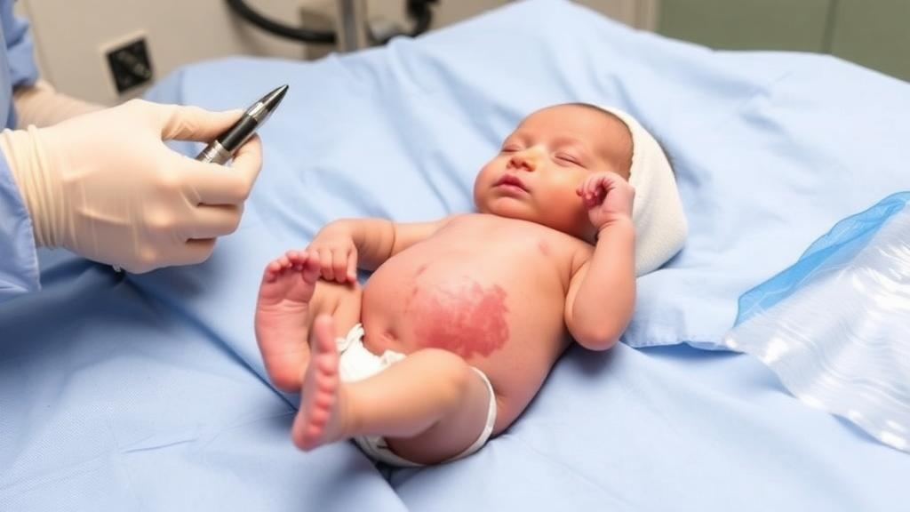
A newborn girl in China who developed life-threatening pulmonary hypertension and active bleeding from a giant congenital hemangioma on her right forearm has made a full recovery after an emergency combination of coil embolization, sclerotherapy, and oral propranolol, according to a case report published in Frontiers in Medicine. The mass, first detected by prenatal ultrasound at 23 weeks of gestation, harbored a high-flow arteriovenous fistula that progressively strained the infant’s circulation after birth. Within days of delivery, the baby was struggling to breathe and the ulcerated surface of the tumor was bleeding. A multidisciplinary team at West China Second University Hospital of Sichuan University performed emergency catheter-based embolization followed by direct injection of sclerosing agents into the lesion, then started beta-blocker therapy. Four weeks after the baseline cardiac examination, follow-up echocardiography documented complete resolution of the pulmonary hypertension, and the infant later achieved all age-appropriate developmental milestones with normal function of the affected arm.
Congenital hemangioma is a rare benign vascular tumor that is present at birth and fully developed, and it differs from infantile hemangioma in both biology and behavior. Clinicians recognize three main subtypes based on natural behavior: rapidly involuting congenital hemangioma, which shrinks quickly after birth; non-involuting congenital hemangioma, which persists indefinitely; and partially involuting congenital hemangioma, which displays characteristics overlapping the other two. Growing genetic evidence indicates that most cases across all three subtypes arise from somatic activating variants in the genes GNAQ and its paralog GNA11, suggesting that they represent one disease category rather than three separate entities. These tumors can appear on the head and neck, trunk, or limbs, and large lesions may drive fetal high-output heart failure, hydrops fetalis, anemia, thrombocytopenia, and even death.
The fistula complicating this case belongs to a different family of vascular disease altogether. Vascular malformations arise from abnormal blood vessel morphogenesis rather than from proliferating tumor cells, and endothelial cell renewal within them remains normal. They are classified by the vessels involved and their flow characteristics: low-flow lesions involve capillaries, veins, lymphatic channels, or mixtures of these, while high-flow lesions involve arteries or direct arteriovenous connections. An arteriovenous malformation is a high-flow malformation composed of abnormal arteries, veins, and capillaries in which blood shunts directly from the arterial to the venous side without passing through a capillary bed. In the trunk and extremities, such lesions typically produce a pulsatile mass, pain, ulceration, bleeding, tissue necrosis, enlarged draining veins, venous hypertension, or heart failure. Because of this high-flow shunting, arteriovenous malformations are more invasive than other congenital vascular malformations and are often accompanied by life- or limb-threatening complications, which is why early and active treatment is generally recommended, even though options remain limited and the disease carries significant morbidity and mortality.
The prenatal course of this case illustrates how such lesions evolve under close surveillance. The fetus was transferred to West China Second University Hospital at 23 weeks of gestation with a progressively enlarging mass on the right forearm, and a multidisciplinary team evaluated the fetus comprehensively while tracking the lesion. At 25 weeks and 3 days, magnetic resonance imaging showed the mass measured approximately 3.48 by 2.02 by 3.55 centimeters, with a multistrip expanding tubular structure and a possibility of hemorrhage, raising suspicion of congenital hemangioma. By 38 weeks, color Doppler ultrasound of the right upper limb showed a mixed-echo mass measuring 6.1 by 7.4 by 8.2 centimeters, suggesting vascular malformation. The baby was delivered by cesarean section at 38 weeks and 2 days, weighing 3,010 grams with perfect Apgar scores of 10 at each of the three assessment points. Physical examination then revealed a 7.0 by 6.5 by 5.0 centimeter mass with clear boundaries, slightly high tension, bluish-purple overlying skin, partial skin breakdown with crusting, and mildly elevated local skin temperature. The authors note that the apparent size difference between prenatal ultrasound and postnatal caliper measurement reflects methodological differences between in-utero imaging through a confined acoustic window and surface measurement, not true tumor shrinkage.
Deterioration came quickly. Two days after birth, the infant developed progressive dyspnea and bleeding from the mass. Echocardiography disclosed severe pulmonary hypertension, with an estimated right ventricular systolic pressure of 52 millimeters of mercury derived from a tricuspid regurgitation peak velocity of 3.4 meters per second using the modified Bernoulli equation, accompanied by bidirectional shunting through the ductus arteriosus. Chest radiography ruled out neonatal respiratory distress syndrome, pneumonia, and pneumothorax, while computed tomography angiography confirmed a giant congenital hemangioma with an arteriovenous fistula. Serial laboratory monitoring revealed a distinctive pattern. On day one, before bleeding began, the platelet count was 182 × 10⁹ per liter, coagulation parameters were normal, and D-dimer was markedly elevated at 6.67 milligrams per liter against a reference value below 0.5, suggesting localized intravascular coagulation within the lesion. On day two, coinciding with active bleeding, platelets fell to 73 × 10⁹ per liter, clotting times showed only minimal prolongation, fibrinogen fell slightly to 2.3 grams per liter but remained normal, and D-dimer rose further to 8.15 milligrams per liter.
The team deliberately excluded Kasabach–Merritt phenomenon, a consumptive coagulopathy classically linked to kaposiform hemangioendothelioma and tufted angioma and only exceedingly rarely reported in congenital hemangioma. The platelet nadir of 73 × 10⁹ per liter never reached the profound thrombocytopenia below 50 × 10⁹ per liter typical of that syndrome; coagulation parameters stayed near-normal with preserved fibrinogen, and the marked D-dimer elevation with normal fibrinogen pointed to localized rather than systemic consumption; the bleeding was confined to the ulcerated skin rather than diffuse; and the underlying tumor type made the syndrome unlikely. Bleeding was therefore attributed primarily to local ulceration and high-flow shunting. Coagulation studies were not repeated after the procedure because clinical bleeding had ceased and the infant was hemodynamically stable, and serial hemoglobin levels remained stable throughout the acute phase without any need for transfusion.
With the team suspecting that the progressive dyspnea stemmed from pulmonary hypertension driven by the giant hemangioma and its fistula, arteriography was performed to confirm the lesion, and emergency combined embolization and sclerotherapy followed immediately. Via right femoral artery and vein puncture with 5-French sheaths placed after systemic heparinization, selective catheterization of the right subclavian artery confirmed a high-flow arteriovenous fistula with dilated, tortuous feeding arteries. A 4F Cobra catheter was advanced into the dilated draining vein, where two 8-millimeter Nester coils were deployed. The operators then punctured the lesional vessels at multiple points through the skin and placed eight additional coils—three 10-millimeter, one 8-millimeter, three 6-millimeter, and one 4-millimeter—for a total of ten coils ranging from 4 to 10 millimeters. Once all coils were in place, a mixture of 0.5 milliliters of iodized oil and 2 milliliters of absolute ethanol was injected, followed by 3 milligrams of bleomycin combined with contrast medium and iodized oil for sclerotherapy. Post-procedural angiography showed reduced lesion opacification and slowed venous drainage, and no immediate procedural complications occurred.
Pharmacotherapy accompanied the intervention. Oral propranolol was prescribed at 1 milligram per kilogram twice daily after contraindications were excluded, then gradually increased to 2 milligrams per kilogram per day. During titration and for 72 hours afterward, continuous telemetry was maintained, with heart rate checked every two hours, blood pressure every four hours, and capillary blood glucose every four hours for 48 hours and then every six hours; all values stayed within age-appropriate ranges, with no bradycardia, hypotension, or hypoglycemia documented. The clinical condition improved, and the mass gradually shrank during follow-up. Baseline echocardiography had shown moderate tricuspid regurgitation with a peak velocity of 3.5 meters per second and a peak gradient of 49 millimeters of mercury, yielding an estimated pulmonary artery systolic pressure of roughly 52 millimeters of mercury, plus trace pulmonary regurgitation from which mean pulmonary artery pressure was estimated at about 35 millimeters of mercury. There was also a 5.2-millimeter patent ductus arteriosus with predominantly right-to-left bidirectional shunting, bidirectional interatrial shunting through two communications of approximately 2 millimeters each, right ventricular wall thickening of 3.1 millimeters, and D-shaped septal motion consistent with right ventricular pressure overload. Follow-up echocardiography four weeks after baseline, fourteen days after embolization, showed normal left ventricular systolic function with an ejection fraction of 66 percent and fractional shortening of 34 percent, normal right heart dimensions, no measurable tricuspid regurgitation jet, no visible ductus, and only a small residual interatrial left-to-right shunt—documenting complete resolution of the pulmonary hypertension. The infant achieved all age-appropriate developmental milestones, with full range of motion, normal grip strength, and no residual edema or compression sequelae in the forearm and hand.
The report situates the case against a sparse literature. Giant congenital hemangiomas of the extremities combined with arteriovenous malformation have rarely been described, and previous accounts include a fetus with a giant congenital hemangioma and arteriovenous malformation that suffered fetal malnutrition, hydrops, and neonatal death, as well as an infant with a giant axillary hemangioma and subclavian arteriovenous fistula who developed heart failure at birth. Among patients with hepatic arteriovenous fistula, more than 50 percent develop heart dysfunction, and 50 to 90 percent die of severe heart failure within the first week of life. A summarized series of 20 congenital hemangioma cases found that 70 percent of affected fetuses survived to birth, with complications such as elevated cardiothoracic ratio, atrioventricular valve regurgitation, and fetal hydrops occurring in 40 percent, and larger lesions—67 percent with maximum diameter of at least 5 centimeters and all with volume of at least 40 cubic centimeters—more likely to cause trouble. In the present case, the authors justified immediate intervention over observation by pointing to active bleeding, a lesion substantially larger than the previously reported axillary case managed conservatively, severe pulmonary hypertension progressing despite respiratory support, and a trajectory of rapid deterioration rather than gradual improvement.
The authors acknowledge important limitations. The diagnosis rested solely on prenatal and postnatal imaging—MRI, Doppler ultrasound, computed tomography angiography, and angiography—because the infant’s unstable condition and the risk of provoking hemorrhage precluded biopsy, so neither histopathological examination nor molecular testing for the characteristic GNAQ and GNA11 mutations was performed. A primary high-flow arteriovenous malformation with secondary mass effect therefore cannot be definitively excluded, and the diagnostic conclusion should be interpreted with caution. The propranolol component also demands careful reading: congenital hemangioma is driven by GNAQ and GNA11 mutations rather than the biology of infantile hemangioma, and some researchers have criticized beta-blocker use for congenital hemangioma, noting that evidence-based support exists only for infantile hemangioma. In this case propranolol was given as an off-label adjunct after emergency embolization and sclerotherapy, based on a multidisciplinary risk–benefit assessment in a life-threatening situation, and the authors stress that a single favorable outcome does not establish efficacy. Their broader message is that clinicians should remain alert to congenital hemangioma with arteriovenous malformation as a cause of severe pulmonary hypertension and bleeding, and that prompt, integrated perinatal management by a multidisciplinary team—reserving intervention for cases with active hemorrhage, progressive hemodynamic compromise, or failed conservative care—can achieve favorable outcomes.
Subject of Research: A neonatal case report of giant congenital hemangioma with arteriovenous fistula treated by embolization, sclerotherapy, and propranolol
Article Title: Case Report: Fetal giant congenital hemangioma with arteriovenous malformation: to be or not to be
Article References: Yue, P., Ma, F., Ma, L., Wan, J., Liu, X., & Hu, F. (2026). Case Report: Fetal giant congenital hemangioma with arteriovenous malformation: to be or not to be. Frontiers in Medicine, 13, Article 1925509. https://doi.org/10.3389/fmed.2026.1925509
Image Credits: AI Generated
DOI: 10.3389/fmed.2026.1925509
Keywords: congenital hemangioma, arteriovenous fistula, arteriovenous malformation, pulmonary hypertension, coil embolization, sclerotherapy, propranolol, neonate, prenatal diagnosis, GNAQ mutation, vascular malformation, case report
News Source: Ophelia Keating. (October 24, 2026). Emergency Embolization Rescues Newborn With Giant Forearm Hemangioma and Arteriovenous Fistula. Scienmag.