Patients with elastofibroma dorsi demonstrated significantly lower lesion attenuation on computed tomography compared to adjacent skeletal muscle in a retrospective, single-centre study. Researchers Atilla Can, Halil Şen, and Hüseyin Yıldıran evaluated patient-level attenuation values to determine whether standardized radiologic density measurements could quantitatively distinguish these benign soft-tissue lesions from normal surrounding muscular tissue.
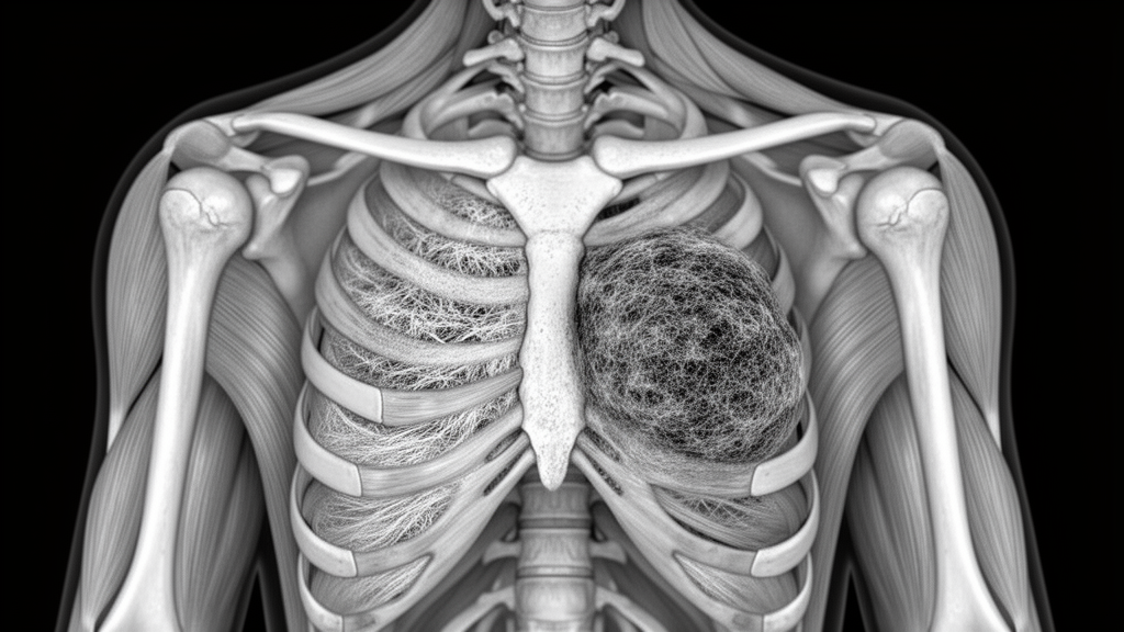
Elastofibroma dorsi represents a benign soft-tissue tumour that characteristically arises within the subscapular region. While the anatomic location and structural appearance of these lesions often provide diagnostic clues, computed tomography attenuation quantified through Hounsfield unit measurements was investigated to supply complementary objective information regarding internal tissue characteristics across individual surgical patients.
The investigated clinical cohort comprised eleven consecutive patients who underwent surgical resection for histopathologically verified elastofibroma dorsi. Among these surgical cases, female patients predominated substantially, accounting for ten individuals or 90.9 percent of the study group, while one patient, representing 9.1 percent, was male. The overall mean age recorded across the surgical cohort was 59.0 ± 11.28 years.
Prior to surgical intervention, symptomatic presentations among the eleven patients included localized pain reported by six individuals, representing 54.5 percent of the cohort. Additionally, five patients, accounting for 45.5 percent of the resected group, presented with clinically palpable swelling at the lesion site before undergoing definitive operative management.
Preoperative computed tomography examinations suitable for standardized Hounsfield unit analysis were available for nine of the eleven surgically treated patients. For individuals presenting with bilateral elastofibroma dorsi, investigators recorded attenuation measurements from both sides and subsequently averaged the right- and left-sided values. This methodology yielded exactly one representative elastofibroma dorsi value and one corresponding adjacent serratus anterior muscle value for each patient.
In the final nine-patient imaging evaluation, the mean computed tomography attenuation recorded for elastofibroma dorsi was 27.89 ± 12.72 Hounsfield units, whereas the adjacent serratus anterior muscle exhibited a higher mean value of 37.89 ± 10.46 Hounsfield units. After confirming the normality of paired differences, a paired-samples t test demonstrated a statistically significant mean paired difference of 10.0 Hounsfield units between the tissues, with a 95 percent confidence interval spanning 4.36 to 15.64 Hounsfield units and a calculated P value of 0.003.
The study authors concluded that elastofibroma dorsi lesions consistently exhibit lower patient-level computed tomography attenuation than adjacent muscular structures, suggesting that quantitative Hounsfield unit analysis can provide useful supportive data when interpreted alongside typical anatomical location and lesion morphology. However, the researchers emphasized key diagnostic limitations, noting that their single-centre investigation did not assess diagnostic specificity against other soft-tissue tumours, nor did it establish a definitive role for attenuation values in preoperative surgical decision-making.
Subject of Research: Computed tomography Hounsfield unit attenuation in elastofibroma dorsi compared with adjacent skeletal muscle
Article Title: Hounsfield unit measurements on computed tomography in elastofibroma dorsi: a patient-level comparison with adjacent skeletal muscle
Article References: Can, A., Şen, H., & Yıldıran, H. (2026). Hounsfield unit measurements on computed tomography in elastofibroma dorsi: a patient-level comparison with adjacent skeletal muscle. Langenbeck's Archives of Surgery. https://doi.org/10.1007/s00423-026-04199-9
Image Credits: AI Generated
DOI: 10.1007/s00423-026-04199-9
Keywords: elastofibroma dorsi, computed tomography, Hounsfield units, attenuation, serratus anterior muscle, subscapular tumour, benign soft-tissue tumour, soft-tissue lesions, retrospective study, paired-samples t test, histopathological confirmation, surgical resection, palpable swelling, preoperative imaging, Atilla Can, Halil Şen, Hüseyin Yıldıran
News Source: Ophelia Keating. (November 18, 2026). Elastofibroma Dorsi Displays Lower Computed Tomography Attenuation Than Adjacent Skeletal Muscle. Scienmag.