Mitral regurgitation, the most common left-sided valvular heart disease, places a substantial burden on healthcare systems worldwide, particularly as the prevalence of symptomatic moderate-to-severe disease rises with advancing age. While surgical repair has long been the standard, transcatheter mitral valve interventions (TMVIs), such as mitral transcatheter edge-to-edge repair (M-TEER) and transcatheter mitral valve replacement (TMVR), have emerged as essential therapeutic alternatives for patients considered to be at high or prohibitive surgical risk. Despite the rapid standardization of these procedures, consensus guidelines have historically provided broad, modality-level imaging recommendations rather than formalizing the distinct requirements of different intervention types. A new state-of-the-art review has addressed this procedural gap by formalizing two fundamentally divergent imaging paradigms: a leaflet-level, temporally driven framework for M-TEER and an annular-level, spatially driven framework for TMVR. This structured approach aims to optimize procedural precision, enhance interdisciplinary communication, and reduce the risk of complications associated with applying a uniform imaging strategy to mechanistically distinct interventions.
The mitral valve apparatus is defined by inherent three-dimensional complexity, including a saddle-shaped annulus, dynamic leaflet excursion across the cardiac cycle, and critical spatial relationships with the left ventricular outflow tract (LVOT) and aortic root. In patients with secondary mitral regurgitation, this structural complexity is often exacerbated by left ventricular remodeling, papillary muscle displacement, and progressive annular dilatation. While fluoroscopy remains an essential tool for catheter navigation and device deployment, its reliance on two-dimensional projections of complex, motion-dependent soft-tissue structures is insufficient for the demands of modern TMVI. As a result, echocardiography has evolved into the primary intraprocedural guidance modality, providing the continuous anatomical visualization and hemodynamic assessment necessary for safe and effective procedural execution. The effectiveness of this echocardiographic guidance, however, depends less on individual modalities and more on their structured integration into a cohesive, workflow-driven architecture that supports continuous spatial orientation throughout each procedural phase.
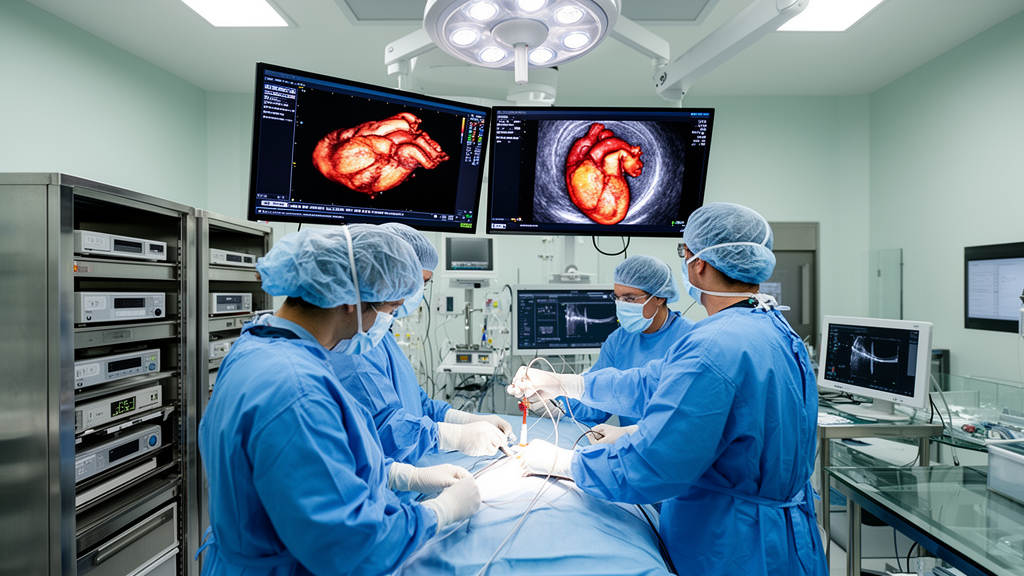
Central to this integrated framework is the use of real-time multiplanar reconstruction (MPR), which converts three-dimensional volumetric echocardiographic datasets into multiple simultaneously displayed orthogonal imaging planes. Unlike sequential two-dimensional imaging, which requires the operator to perform cognitive spatial reconstruction—a process prone to error in challenging anatomical environments—real-time MPR maintains true orthogonality relative to key anatomical landmarks regardless of probe position. This geometric stability provides a consistent spatial reference frame, which is critical for transseptal puncture and subsequent navigation within the left atrium. By bridging the temporal precision of two-dimensional transesophageal echocardiography (TEE) with the spatial clarity of three-dimensional acquisitions, MPR functions as the geometric backbone of modern structural heart echocardiography. The ability to visualize the puncture site, septal thickness, and trajectory in a single, stable view minimizes the reliance on probe-dependent estimation and enables more reproducible procedural maneuvers across diverse device platforms.
Echocardiography-fluoroscopy fusion imaging further enhances this spatial integration by co-registering real-time echocardiographic data with fluoroscopic projections in a shared coordinate system. By projecting soft-tissue landmarks directly onto the fluoroscopic field, this technique reduces the cognitive burden associated with translating information between spatially independent displays. While fusion imaging is highly valuable during catheter advancement, device orientation, and final positional confirmation, researchers emphasize that it should be viewed as an adjunctive spatial integration tool rather than a replacement for primary echocardiographic guidance. Its reliability is critically dependent on the accuracy of co-registration and the maintenance of that alignment throughout the procedure, as drift caused by patient movement or transducer repositioning can degrade spatial accuracy. As a result, the technique is best utilized as a functional bridge toward fully integrated, platform-based guidance, particularly in procedures where fine positional adjustments are required.
For patients in whom transesophageal echocardiography is not feasible or desirable, such as those with esophageal pathology or those preferring conscious sedation, three-dimensional intracardiac echocardiography (ICE) has emerged as a patient-selective imaging option. Advanced catheter-mounted matrix-array transducers enable real-time volumetric intracardiac visualization, expanding procedural accessibility for high-risk, elderly, or frail candidates. However, the field of view is constrained by the proximity of the catheter to the target anatomy, and the current evidence base for its use in TMVI remains limited to case reports and single-center series. Importantly, 3D ICE does not offer the same comprehensive anatomical context or geometric consistency as MPR-based transesophageal imaging, particularly regarding en face visualization of the global mitral apparatus. Consequently, it is best conceptualized as a complementary modality that expands access to care rather than a complete replacement for standard TEE-based workflows.
The role of artificial intelligence (AI)-assisted decision-support tools represents a conceptual shift toward integrating computational layers into intraprocedural workflows. These tools do not generate new anatomical information; instead, they operate on existing three-dimensional TEE volumes, MPR spatial frameworks, and fusion-registered data to automate landmark tracking and provide real-time feedback on device position relative to predefined anatomical targets. By organizing and stabilizing these data, AI-assisted tools help preserve spatial consistency during prolonged procedures or multi-device interventions. This redistribution of cognitive load—where AI manages repetitive spatial monitoring—allows echocardiographers and interventional operators to focus on higher-order clinical decision-making. Emerging clinical data suggest that these tools can enhance team communication and procedural reproducibility, though multicenter prospective studies are still required to define their independent impact on safety and long-term outcomes.
The formal distinction between the imaging requirements for M-TEER and TMVR is crucial for tailoring these workflows effectively. M-TEER focuses on leaflet-level capture within a confined left atrial workspace; therefore, its imaging requirements prioritize high temporal resolution to capture rapid leaflet excursion and facilitate precise orientation relative to the coaptation line. In contrast, TMVR is an annular-level procedure involving large, stiff delivery systems that significantly alter LVOT geometry. The primary imaging priorities for TMVR revolve around accurate spatial navigation, coaxial alignment with the annular plane, and the maintenance of neo-LVOT patency. Failure to distinguish these priorities can lead to procedural risks, such as incomplete leaflet engagement in M-TEER or improper prosthesis seating and LVOT obstruction in TMVR. By tailoring imaging protocols to these specific mechanisms, procedural teams can ensure more effective engagement and improved functional outcomes.
Clinical case examples highlight the utility of this structured approach in complex scenarios. For instance, in an elderly patient with degenerative mitral regurgitation involving the full coaptation line, a multi-device M-TEER strategy can carry a meaningful risk of residual regurgitation that iterative MPR reassessment is essential to manage. Similarly, in TMVR procedures for patients with mitral annular calcification, the inherent irregularity of the annulus often renders traditional fluoroscopic landmarks unreliable. In these instances, MPR-centered spatial control provides the primary echocardiographic reference for annular-level decision-making, ensuring that the prosthesis is correctly positioned and seated even when fluoroscopic visualization is limited. These scenarios underscore the necessity of moving beyond modality-specific expertise toward proficiency in integrated, platform-based imaging environments.
The transition toward integrated, platform-based guidance presents several practical challenges, including resource availability, procedural costs, and institutional expertise disparities. Implementation of advanced imaging platforms requires not only investment in hardware but also a significant evolution in training pathways for both interventional operators and structural echocardiographers. Future developments, such as the integration of augmented reality overlay systems and real-time finite element modeling, promise to further reduce the cognitive burden of spatial translation and enable predictive assessment of outcomes before device deployment. However, these technologies are still in early evaluation and remain technically demanding, requiring rigorous validation across the anatomical heterogeneity characteristic of the TMVI patient population.
Ultimately, the evolution of intraprocedural imaging from a collection of complementary tools into a cohesive, echocardiography-centered platform represents a definitive trajectory for modern structural heart practice. The effectiveness of this architecture depends on structured integration rather than the application of individual modalities. As device platforms continue to diversify and patient cohorts become increasingly complex, the role of intraprocedural imaging as a central determinant of procedural strategy, safety, and scalability will only intensify. Future research must prioritize standardized protocols, prospective multicenter validation, and training frameworks that emphasize the multimodal nature of contemporary structural interventions. Through these advancements, the field aims to move toward a paradigm where procedural success is supported by robust, integrated, and objective-driven guidance across the entire spectrum of mitral valve therapies.
Subject of Research: Echocardiographic guidance frameworks for transcatheter mitral valve interventions
Article Title: Intraprocedural echocardiographic guidance in transcatheter mitral valve interventions: from procedure-specific workflows to integrated multimodal platforms
Article References: Benjamins, S. A., Vrijkorte, M. G. H., Timmers, L., Rensing, B. J. W. M., & Swaans, M. J. (2026). Intraprocedural echocardiographic guidance in transcatheter mitral valve interventions: from procedure-specific workflows to integrated multimodal platforms. Journal of Cardiovascular Imaging, 34(1), Article 31. https://doi.org/10.1186/s44348-026-00092-7
Image Credits: AI Generated
DOI: 10.1186/s44348-026-00092-7
Keywords: Transcatheter Mitral Valve Intervention, Mitral Regurgitation, Echocardiography, Multiplanar Reconstruction, M-TEER, TMVR, Structural Heart Disease, Intraprocedural Guidance, Artificial Intelligence, Fusion Imaging, 3D TEE, 3D ICE, Mitral Annular Calcification, Procedural Workflow, Clinical Imaging, Cardiovascular Imaging, Left Ventricular Outflow Tract, Device Deployment
News Source: Ophelia Keating. (November 18, 2026). New Framework Refines Echocardiographic Guidance for Complex Transcatheter Mitral Valve Procedures. Scienmag.