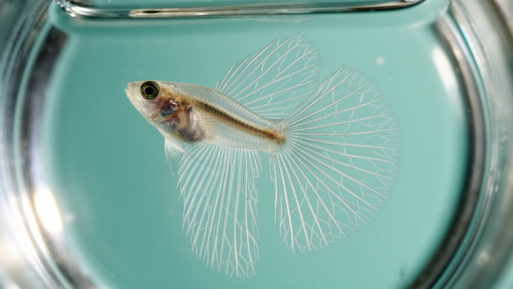
Exposure to low-frequency mechanical vibration selectively disrupts the skeletal development of zebrafish larvae depending on the exact timing of the stimulus, uncovering distinct developmental windows where immature skeletal tissues react to physical forces. In an experimental investigation published in Development Genes and Evolution, researchers Shirine Jeradi and Tamara A. Franz-Odendaal demonstrated that whole-body vibration at 20 Hz altered the morphology of specific caudal fin elements in wild-type AB zebrafish. The findings show that mechanical sensitivity in the vertebrate skeleton is not limited to differentiated, mature bones that regularly endure load-bearing stresses, but is also present in early chondrogenic progenitor cells and notochordal sheath cells prior to terminal differentiation.
Vertebrate skeletons adapt to physical constraints through mechanosensation, yet the cellular mechanisms guiding how immature, developing skeletal elements respond to mechanical signals remain incompletely understood. Vertebrate bones develop primarily through two conventional pathways: intramembranous ossification, where mature osteoblasts directly deposit bone matrix, and endochondral ossification, where chondroblasts first establish a cartilaginous template that osteoblasts subsequently invade and mineralize. In certain fishes, including teleosts such as zebrafish, an additional pathway termed perichordal ossification forms vertebral structures without an intermediate cartilage stage and initiates independently of osteoblasts. In zebrafish larvae, vertebral precursors called chordacentra form through direct mineralization of the notochord sheath via collagen matrix deposition by chordoblasts, providing a distinct comparative system alongside standard cartilaginous condensation events.
To investigate how controlled vibrational loading influences these diverse ossification modes across embryonic and larval growth, the research team examined wild-type zebrafish maintained at 28.5 degrees Celsius under approved institutional animal care protocols. Healthy larvae were systematically separated into experimental cohorts and placed in petri dishes holding 35 milliliters of fish water atop a custom-built vibration apparatus. This platform incorporated four parallel-connected speakers positioned inside an incubator and linked to a frequency controller and timing circuit. Building on earlier pilot evaluations testing sinusoidal vibration waves at frequencies of 8 to 10 Hz and 20 Hz, the authors designated 20 Hz as an active mechanical influencer for their experimental schedule. Cohorts of twenty embryos or larvae were subjected either to a two-hour vibration exposure starting at the bud stage, approximately 10 hours post-fertilization, or to four-day exposures commencing at 2, 3, 4, or 5 days post-fertilization.
Following vibrational exposure, fish were maintained in rearing containers until reaching a standard length endpoint between 5.5 and 6.5 millimeters, corresponding to approximately 4 to 6 weeks post-fertilization, before undergoing euthanasia and fixation in 4 percent paraformaldehyde. A subset of specimens exposed from 5 to 9 days post-fertilization was fixed at 9 days post-fertilization for histological and molecular assays. To prevent the acidic degradation of small mineralized elements, the investigators applied an acid-free dual-staining procedure utilizing Alcian blue for cartilage and Alizarin red for mineralized bone, modified from Kimmel and Walker. The stained skeletons were evaluated across multiple anatomical regions, including Meckel’s cartilage, palatoquadrate, ethmoid plate, branchial arches, jaw structures, fin rays, and notochordal centra, with particular emphasis placed on posterior axial caudal elements such as ural centra 1 through 3, preural vertebrae, parhypural elements, and hypurals 1 through 5.
The experimental observations demonstrated a striking age-dependent and bone-specific susceptibility to whole-body vibration. Ural centrum 3, an element that ossifies through the perichordal pathway without passing through a cartilaginous precursor, showed complete developmental impairment in all eighteen larvae subjected to 20 Hz vibration for two hours starting at the bud stage. In contrast, unexposed control larvae showed zero defects across eighteen individuals. When four-day vibrational treatments commenced at 2 days post-fertilization, ural centrum 3 development failed in 37.5 percent of specimens, representing 3 out of 8 larvae. Exposure starting at 3 days post-fertilization resulted in defects in 18.18 percent, or 2 out of 11 larvae, while exposure starting at 4 days post-fertilization yielded defects in 10 percent, or 1 out of 10 larvae. Exposure delayed until 5 days post-fertilization produced no ural centrum 3 defects among 22 evaluated larvae, matching the baseline observed in controls.
Remarkably, this sensitivity was confined exclusively to ural centrum 3 within the perichordal series. The remaining vertebral centra developed normally across all treated age groups, with standard-length counts showing no statistically significant variation in centra-derived vertebrae relative to unexposed controls. The authors noted that while most chordacentra undergo initial sheath mineralization followed by autocentral ossification involving osteoblasts, ural centrum 3 does not follow this conventional trajectory and lacks osteoblast colonization up to 8.3 millimeters standard length. Because osteoblasts serve as primary mechanosensors in anosteocytic bones, the distinct cell populations forming ural centrum 3 may explain why presumptive notochordal sheath cells in this single segment display heightened mechanical vulnerability during initial embryogenesis before progressively losing sensitivity as development advances.
Conversely, the cartilaginous elements of the caudal fin endoskeleton exhibited the reverse temporal pattern of vulnerability. Exposure to 20 Hz vibration initiated at the bud stage or at 2 or 3 days post-fertilization caused no structural defects in caudal fin cartilages, with all evaluated larvae exhibiting normal parhypural and hypural morphology. However, when four-day vibration exposures were initiated at 4 days post-fertilization, 30 percent of larvae, or 3 out of 10 specimens, developed patterning defects consisting of discontinuous hypural 1 structures or ectopic fusions uniting the parhypural and hypural 1 elements. When four-day treatments began at 5 days post-fertilization, ectopic fusions connecting the parhypural and hypural 1 appeared in 36.4 percent of specimens, or 8 out of 22 larvae. Other skeletal structures, including craniofacial jaw cartilages, opercular bones, and fin rays, remained completely unaltered, demonstrating anatomical specificity.
To elucidate the molecular pathways mediating these caudal cartilage disruptions, the researchers examined early markers of chondrogenesis. Because the transcription factor Sox9 orchestrates chondrogenic condensation and activates downstream structural genes like collagen type II alpha 1, the authors performed whole-mount in situ hybridization targeting both zebrafish co-orthologues, sox9a and sox9b, in 9 days post-fertilization larvae following exposure from 5 to 9 days post-fertilization. Untreated controls displayed discrete caudal cell clusters expressing sox9a in 10 out of 10 specimens and sox9b in 7 out of 7 specimens. Following vibration exposure, these two distinct expression clusters were fully maintained in 10 out of 10 larvae for sox9a and 12 out of 12 larvae for sox9b, demonstrating that vibration does not suppress the transcriptional commitment of mesenchymal progenitors to a chondroblastic fate. Histochemical staining for alkaline phosphatase, an early indicator of skeletal mineralization capacity, also remained present and properly organized across all twenty evaluated vibration-exposed larvae, matching fourteen untreated controls.
Because the cartilage defects occurred without altering Sox9 gene expression or alkaline phosphatase activity, the researchers conducted quantitative morphometric measurements of the early chondrogenic condensations in 9 days post-fertilization larvae. Three discrete condensations were mapped in the ventral median fin fold: condensation 3 rostrally, condensation 2 intermediately, and condensation 1 caudally, corresponding respectively to the presumptive parhypural, hypural 1, and hypural 2 elements. Systemic measurements revealed that the average physical distance separating condensations 2 and 3 was significantly reduced in vibration-exposed larvae compared to controls, with statistical significance exceeding p less than 0.01 across cohorts of 22 exposed and 19 control larvae. In contrast, the distance between condensations 1 and 2 showed no statistically significant change. Direct area measurements showed that individual condensation sizes were statistically indistinguishable between exposed and control fish, verifying that the spatial proximity between condensations 2 and 3 did not arise from cellular swelling or expanded condensation boundaries.
These experimental measurements indicate that low-frequency mechanical vibration directly alters the physical spacing between adjacent chondrogenic condensations during initial fin fold patterning, predisposing neighboring cartilages to ectopic fusion during subsequent growth. The differential vulnerability of parhypural and hypural 1 compared to hypurals 2 through 5 aligns with the natural developmental chronology of the zebrafish caudal endoskeleton, in which the lower lobe elements condense between 3.8 and 4.2 millimeters standard length while the upper hypurals form later between 4.4 and 5.0 millimeters standard length. Because the treatments ending at 8 and 9 days post-fertilization overlapped precisely with the emergence of the parhypural and hypural 1 condensations, those early mesenchymal aggregates bore the brunt of the vibrational loading. The findings confirm that uncommitted and condensing skeletal progenitor cells possess intrinsic mechanosensitive capacities in vivo, operating independently of the classic Sox9 cascade to influence skeletal morphology.
Subject of Research: Mechanical stimulus effects on zebrafish skeletal development
Article Title: Vibration exposure uncovers a critical early developmental window for zebrafish caudal fin development
Article References: Jeradi, S., & Franz-Odendaal, T. A. (2022). Vibration exposure uncovers a critical early developmental window for zebrafish caudal fin development. Development Genes and Evolution, 232(2-4), 67-79. https://doi.org/10.1007/s00427-022-00691-6
Image Credits: AI Generated
DOI: 10.1007/s00427-022-00691-6
Keywords: zebrafish, developmental biology, mechanosensation, skeletal patterning, chondrogenesis, whole-body vibration, caudal fin, osteogenesis, Vibration, exposure, uncovers, critical
News Source: Drew Townsend. (November 11, 2026). Early Vibration Exposure Disrupts Skeletal Patterning in Developing Zebrafish Tail Fin. Scienmag.