
Reston, VA (May 9, 2025)—In a remarkable stride forward for nuclear medicine and molecular imaging, a series of groundbreaking studies have been published ahead of print in the prestigious Journal of Nuclear Medicine (JNM), shedding new light on innovations that promise to revolutionize diagnostics and therapeutics in neuroscience, oncology, and precision health. Published by the Society of Nuclear Medicine and Molecular Imaging (SNMMI), these reports emphasize the cutting-edge progress in developing novel PET tracers, refining imaging techniques, and expanding our understanding of molecular targets critical to disease mechanisms and treatment personalization.
One of the most compelling advancements involves a newly developed positron emission tomography (PET) radioligand known as ^11C-ZTP-1, which specifically targets phosphodiesterase 4B (PDE4B) — an enzyme intricately connected to neuroinflammation and various neuropsychiatric disorders. The ability of ^11C-ZTP-1 to selectively image PDE4B was rigorously validated in preclinical models including rats and non-human primates. This short-lived radiotracer’s unique properties facilitate the possibility of multiple scans within the same day, an innovation that could accelerate both fundamental brain research and the clinical development pipeline for novel neurotherapeutics by providing dynamic, temporal mapping of enzyme activity in vivo.
Expanding our understanding of breast cancer imaging, another pioneering study challenges the traditional categorization of “false positives” in PET imaging by utilizing ^89Zr-labeled antibodies. This approach has demonstrated the capacity to detect HER2-low breast cancer lesions, a subset formerly misclassified and overlooked due to limitations of earlier imaging modalities. This revelation not only broadens the diagnostic scope of HER2 PET imaging but also introduces the potential for identifying patients who could benefit from emerging HER2-targeted therapies, representing a paradigm shift in oncological precision medicine that prioritizes molecular heterogeneity.
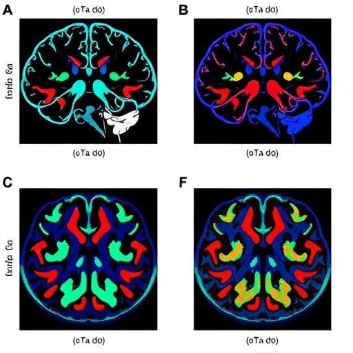
Further enhancements in brain imaging are demonstrated by the introduction of ^18F-K-40, a novel PET tracer that permits visualization of AMPA receptors in living human subjects. AMPA receptors, fundamental to excitatory neurotransmission and synaptic plasticity, play a pivotal role in cognitive processes and neurological health. By matching the specificity and sensitivity of prior tracers with the added advantage of a longer half-life, ^18F-K-40 enables more flexible and accessible imaging protocols, promising to deepen investigations into neurological and psychiatric diseases where AMPA receptor dysfunction is implicated, such as epilepsy, depression, and neurodegeneration.
In a separate investigation, whole-body PET imaging using ^11C-carfentanil, a selective agonist for μ-opioid receptors, has unveiled significant sex-based differences in receptor distribution and naloxone-mediated receptor blockade within the central nervous system. By capturing the nuanced neurobiological variations between men and women in brain regions associated with pain modulation and addiction, this study provides critical insights that could inform sex-specific strategies for managing opioid use disorder and improving the efficacy of analgesic therapies. Understanding these distinctions enhances our grasp of opioid pharmacodynamics and may lead to more personalized approaches in pain medicine and addiction treatment.
Parallel to these empirical studies, a comprehensive review articulates the expanding role of molecular imaging in human phenomics—the systemic study of phenotypes at a complexity scale. Integrating molecular imaging with multiomics datasets and artificial intelligence (AI), this research underscores the transformative potential of such synergy to offer quantitative, predictive insights. This systems-level approach moves beyond traditional diagnostics, fostering preclinical intervention strategies and facilitating a precision health framework centered on individualized biological complexity and continuous monitoring.
The frontiers of personalized cancer imaging and therapy are also pushed forward through investigations targeting the gastrin-releasing peptide receptor (GRPR). Recognized for its overexpression in multiple tumors, GRPR stands as a promising biomarker and therapeutic target. The transition from bench to bedside is explored, emphasizing the dual role of GRPR-based molecular imaging in both diagnosing diverse cancers and delivering targeted radionuclide therapy. This approach embodies the principles of theranostics, thereby optimizing patient selection and therapeutic efficacy while minimizing off-target effects.
Collectively, these studies exemplify a multifaceted advancement of nuclear medicine, leveraging sophisticated tracer chemistry, enhanced imaging techniques, and system biology approaches to drive forward precision medicine. The implications extend from improved brain disorder management and cancer therapeutics to refined diagnostic accuracy, establishing new standards for molecular imaging’s integration into clinical workflows. These innovations hint at a future where individualized treatment selection and comprehensive phenomic assessment are hallmarks of patient care.
Moreover, these pioneering PET imaging tools highlight a key shift toward creating tracers tailored not only for improved diagnostic clarity but also for enabling dynamic therapeutic monitoring. The ability to visualize enzymatic activity, receptor binding, and cellular heterogeneity in vivo opens unparalleled avenues for drug development, patient stratification, and real-time treatment assessment, cultivating an era of truly personalized medicine underpinned by actionable molecular insights.
Advancements in whole-body imaging, such as those achieved with ^11C-carfentanil, demonstrate the feasibility of moving beyond regional brain studies to systems-level interrogation of receptor distributions and pharmacokinetics. This approach facilitates a holistic understanding of complex biological systems and sexes differences, which are often underappreciated in neuropharmacology—offering fertile ground for new discoveries that properly integrate biological diversity into therapeutic design.
The integration of artificial intelligence and multiomics into molecular imaging reveals an exciting interdisciplinary frontier, exploiting the vast data generated through imaging technologies to create predictive models and refined diagnostics. This bidirectional relationship harnesses AI’s capacity to decode complex imaging patterns, while molecular imaging provides the spatially and temporally dense data necessary for nuanced model training—a symbiosis poised to revolutionize predictive medicine, early disease detection, and personalized intervention strategies.
As the field advances, the translational pipeline for molecular imaging agents such as those targeting PDE4B and GRPR is essential for transforming laboratory discoveries into clinical realities. Accelerating regulatory approval, expanding accessibility, and ensuring cost-effectiveness remain challenges, yet the potential impact on patient diagnosis, treatment personalization, and outcome prediction affirms the immense value of these innovations for modern healthcare systems.
The Journal of Nuclear Medicine continues to be at the forefront of disseminating pivotal research that redefines our approach to molecular diagnostics and therapeutics. With a global audience exceeding 15 million annual accesses, the journal serves as an indispensable resource for clinicians, researchers, and industry stakeholders dedicated to harnessing nuclear medicine’s potential to improve patient lives.
For the latest in molecular imaging advancements, practitioners and researchers are encouraged to explore JNM’s comprehensive coverage as the field moves toward increasingly sophisticated, integrative, and patient-specific methodologies that stand to reshape the landscape of healthcare.
Subject of Research: Molecular imaging innovations in neuroscience, oncology, and precision health
Article Title: Various – including “New Brain Imaging Tool Targets Key Enzyme in Mental Health” and “Targeting GRPR: A New Frontier in Personalized Cancer Imaging and Therapy”
News Publication Date: May 9, 2025
Web References:
https://doi.org/10.2967/jnumed.124.269159
https://doi.org/10.2967/jnumed.124.269227
https://doi.org/10.2967/jnumed.124.269405
https://doi.org/10.2967/jnumed.124.269413
https://doi.org/10.2967/jnumed.124.267660
https://doi.org/10.2967/jnumed.124.269444
Keywords: Molecular imaging, PET tracers, PDE4B, HER2-low breast cancer, AMPA receptors, μ-opioid receptors, GRPR, precision medicine, theranostics, neuropsychiatric disorders, molecular phenomics, sex differences, multiomics, artificial intelligence
Tags: breast cancer imaging techniquesdynamic enzyme activity mappingJournal of Nuclear Medicine highlightsmolecular imaging innovationsneuroinflammation researchneuropsychiatric disorder diagnosticsnuclear medicine advancementsPET tracer developmentphosphodiesterase 4B imagingprecision health advancementspreclinical validation studiesSociety of Nuclear Medicine and Molecular Imaging

