One of the most remarkable feats in human biology happens quietly in the first weeks of pregnancy, when a handful of embryo-derived cells begin constructing the placenta, the temporary organ that will deliver oxygen, nutrients and immune protection to a growing fetus. A new review published in Molecular Biology Reports argues that a single adhesion molecule, E-cadherin, acts as a dynamic molecular switch governing how those cells choose their fates. Rather than serving as a static glue that simply holds tissue together, E-cadherin rises and falls in carefully choreographed waves across different trophoblast subpopulations, and the timing of those waves appears to determine whether cells fuse into the placenta’s exchange barrier or break away to invade the mother’s uterus.
The review, authored by Enoch Appiah Adu-Gyamfi and Ethan Backes of the University of Wisconsin-Stout, synthesizes decades of fragmented literature into a unified framework. The authors contend that human placentation depends on the precise differentiation of trophoblasts, the specialized cells of the outer embryo, into distinct lineages, and that this differentiation is strictly regulated by the spatio-temporal expression of key genes. E-cadherin, a 120-kilodalton transmembrane glycoprotein encoded by the CDH1 gene, sits at the center of that regulatory story. When its expression pattern goes wrong, the consequences can be severe: dysregulation is evident in placentas from pregnancies complicated by preeclampsia and fetal growth restriction, two of the most common and dangerous obstetric complications.
To understand why a single adhesion protein matters so much, it helps to grasp the scale of the construction project. After the blastocyst adheres to the uterine lining, the trophectoderm gives rise to cytotrophoblasts, mononuclear and actively proliferating cells that function as stem cells for every other trophoblast lineage. These cells follow two main routes. Along the villous pathway, cytotrophoblasts multiply and fuse with one another to form the syncytiotrophoblast, a multinucleated layer that serves as the barrier controlling physiological exchange between mother and fetus. Along the extravillous pathway, they differentiate into highly invasive extravillous trophoblasts that anchor the placenta to the uterus and remodel maternal blood vessels to feed the growing fetus.
The extravillous journey is extraordinary in its own right. Proximal column trophoblasts, which remain near the villous tree, give rise to distal column trophoblasts that migrate into the decidua and become interstitial extravillous trophoblasts. Some of these invade and open the uterine glands, ensuring a supply of nutrient-rich secretions before full maternal blood flow begins. Others form temporary plugs inside the spiral arteries, deliberately restricting blood flow to maintain the low-oxygen environment that early placental development requires. Later, as fetal demands grow, endovascular trophoblasts dismantle the muscular walls of those arteries and migrate backward up their lumens, replacing the maternal endothelial lining. The arteries lose their contractility and transform from narrow, high-resistance coils into wide, flaccid conduits capable of delivering large volumes of blood at low pressure, a system fully established by the twelfth week of gestation.
E-cadherin’s structure explains both its adhesive power and its signaling reach. Its extracellular region consists of five tandemly repeated domains that form zipper-like, calcium-dependent bonds with E-cadherin molecules on neighboring cells. Its intracellular tail anchors the protein to the actin cytoskeleton through a complex of catenin proteins, linking cell-cell adhesion directly to the cell’s mechanical and signaling machinery. Beyond holding tissue together, E-cadherin regulates apico-basal polarity, enforces contact inhibition of proliferation by sequestering beta-catenin at the membrane, and is classified as a potent tumor suppressor. When cancer cells initiate epithelial-to-mesenchymal transition, transcriptional reprogramming shuts down E-cadherin, dismantling the adhesion complex and freeing cells to detach and migrate. Placental biology, the review argues, co-opts this same machinery for constructive rather than destructive ends.
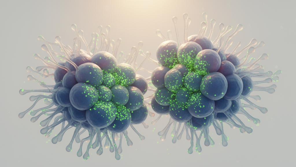
Along the villous pathway, the evidence reveals a striking rise-and-fall pattern. Pioneering work by C. Coutifaris and colleagues showed that E-cadherin accumulates on the surfaces of cytotrophoblasts at points of cell-cell contact, peaking around twenty-four hours as cells aggregate into clusters. But once fusion begins, E-cadherin must disappear. Follow-up studies demonstrated that its loss is a requirement, not merely a consequence, of trophoblast fusion: high levels physically aggregate the mononuclear cells, and only their dynamic downregulation allows adjacent membranes to dissolve so cytoplasm can merge. In BeWo cell models treated with cAMP analogs or forskolin, fusion coincided with E-cadherin vanishing from cell surfaces, and the transcription factor Twist, which localizes to syncytiotrophoblast nuclei, was shown to repress the protein during this transition.
The review highlights three converging mechanisms that drive E-cadherin down during syncytialization. First, transcriptional repression by Twist, and possibly Snail, silences CDH1 expression. Second, enzymes do the cutting: HtrA4, a protease highly expressed in the placenta, is essential, because when it is silenced or deleted, E-cadherin persists and fusion is completely blocked. ADAM12, a member of the disintegrin and metalloprotease family, potentiates fusion by shedding the E-cadherin ectodomain, destabilizing existing adherens junctions and catalyzing membrane coalescence. Third, and perhaps most surprisingly, physics joins in. Mechanical stress patterns predicted by computational models of villous morphogenesis, when applied to cytotrophoblast monolayers, were shown to facilitate E-cadherin downregulation, with equibiaxial compressive stresses potentially lowering the energetic barrier for membrane fusion by rearranging lipids and proteins at the fusion interface.
Along the extravillous pathway, E-cadherin plays an equally dramatic but opposite-seeming role. Its transient downregulation characterizes a physiological epithelial-to-mesenchymal transition that permits trophoblasts to detach from anchoring columns and invade the decidua. The functional evidence is compelling: when researchers experimentally restored E-cadherin in invasive trophoblasts, the cells lost their migratory shapes, reverted to cohesive aggregates, and exhibited robust contact-dependent inhibition of movement. Repressors such as Twist, Snail, ZEB1 and ZEB2 correlate inversely with E-cadherin in these cells, and the concurrent upregulation of N-cadherin actively promotes the disassembly of E-cadherin-mediated adhesions. Yet the story does not end with invasion. When trophoblasts reach the spiral arteries, E-cadherin expression rises again, cooperating with VE-cadherin and VCAM-1 in a form of vascular mimicry that lets the cells withstand hemodynamic forces and integrate into the vessel wall. This partial reversal of EMT suggests that E-cadherin loss is a transient state, not a terminal commitment.
The clinical stakes are considerable. Persistent E-cadherin expression on the syncytiotrophoblast layer signals incomplete fusion, and its abnormal persistence correlates with preeclampsia and fetal growth restriction, conditions linked to poorly formed syncytium and shallow trophoblast invasion. The review proposes that soluble E-cadherin fragments shed by ADAM12 might be detectable in maternal blood during the first trimester, offering a potential early biomarker of defective placentation, though large-scale translational studies are needed to establish feasibility. Histologically, the disappearance of E-cadherin-stained cell boundaries already serves as a reliable indicator of successful syncytial maturation, and quantifying E-cadherin levels provides a valid, if complementary, experimental metric of fusion efficiency.
What excites the field most, however, is what remains unknown. The authors argue that research must now move from descriptive profiling to functional mechanism. Key questions include whether HtrA4, ADAM12 and compressive forces operate in a linear cascade or parallel networks, whether mechanosensitive channels such as PIEZO1 translate mechanical strain into the intracellular cAMP signals that initiate fusion, and how the intracellular pool of E-cadherin is cleared through endocytosis, proteasomal or lysosomal degradation. Whether trophoblast-derived soluble E-cadherin fragments act as signaling molecules, as they do in cancer, or as decoys disrupting maternal junctions is unexplored. The reviewers call for spatial transcriptomics, single-cell RNA sequencing, atomic force microscopy and advanced membrane imaging to map how tissue stiffness, fluid shear stress and local immune cells, including uterine natural killer cells, trigger the E-cadherin switch cell by cell. If those efforts succeed, a protein once known only as epithelial glue may emerge as both a diagnostic window into pregnancy complications and a therapeutic target in placental disease.
Subject of Research: The role of spatio-temporal E-cadherin expression in regulating trophoblast lineage differentiation during human placentation
Article Title: Spatio-temporal expression of E-cadherin mediates trophoblast lineage transitions during human placentation
Article References: Adu-Gyamfi, E. A., & Backes, E. (2026). Spatio-temporal expression of E-cadherin mediates trophoblast lineage transitions during human placentation. Molecular Biology Reports, 53(1), Article 1678. https://doi.org/10.1007/s11033-026-12837-0
Image Credits: AI Generated
DOI: 10.1007/s11033-026-12837-0
Keywords: E-cadherin, trophoblast, placentation, syncytiotrophoblast, extravillous trophoblast, epithelial-to-mesenchymal transition, preeclampsia, fetal growth restriction, spiral artery remodeling, HtrA4, ADAM12, mechanobiology
News Source: Juliet Wilcox. (October 8, 2026). E-cadherin’s Rising and Falling Levels Steer How the Placenta Builds Itself. Scienmag.