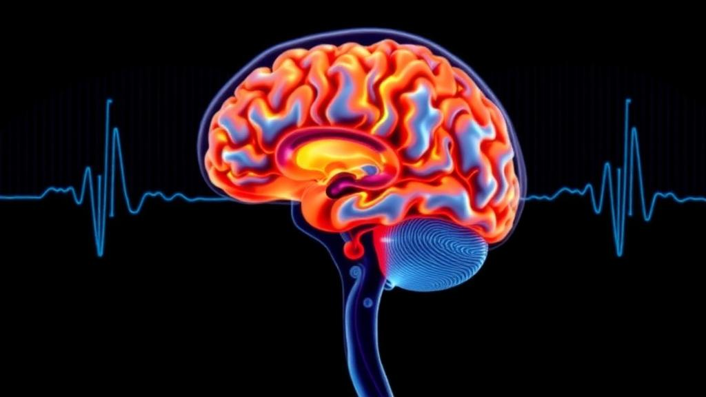
Some of the most dangerous brain injuries are the ones that never show up on a scan. Every year, more than 50 million people worldwide sustain a traumatic brain injury, and countless others experience silent strokes or subclinical damage that leave no visible trace on computed tomography or structural magnetic resonance imaging. Yet inside the skull, something has already changed: neurons fire out of rhythm, synapses lose efficiency, and the brain’s vast communication networks begin to drift. A new technical review published in Frontiers in Medical Technology argues that the key to catching these invisible injuries lies not in sharper pictures of brain structure, but in listening to how the brain talks to itself.
The review, led by Hongmei Ruan and colleagues at Shaoguan University Affiliated Hospital and Gannan Medical University in China, tackles a stubborn clinical problem. Conventional imaging excels at spotting haemorrhage, oedema and infarction, but its detection rate for micro-injuries at the level of neuronal electrophysiology and brain network function is extremely low. Patients with mild traumatic brain injury, or mTBI, frequently walk out of emergency departments with clean scans and persistent headaches, cognitive fatigue and sleep disturbances that are easily overlooked. Without timely intervention, some develop chronic post-concussion symptoms, cognitive decline, anxiety and depression, while those with silent cerebrovascular damage face elevated risks of dementia and stroke.
The authors propose a framework they call Dynamic-Spectral Coupling, or DSC, built on a testable mechanistic hypothesis rather than an established causal model. Its core idea is that local disruptions in the balance between cortical excitation and inhibition, mediated by glutamatergic and GABAergic neurons, may serve as an upstream mechanism driving abnormal switching of whole-brain dynamic network states. When inhibitory neurons weaken, cortical neurons discharge abnormally in synchrony, energy metabolism falters, and synaptic transmission efficiency drops. At the EEG level, this manifests as spectral slowing, with increased low-frequency delta and theta power and reduced high-frequency beta and gamma activity. At the network level, it appears as declining stability in the default mode, salience and frontoparietal networks, characterised by increased switching rates and shortened state dwell times.
A particularly important technical contribution of the review is its treatment of the 1/f aperiodic component of the EEG power spectrum. Unlike periodic oscillations, which reflect rhythmic firing, the aperiodic background signal reflects the overall excitability of neuronal populations. Using parametric modelling frameworks such as FOOOF, researchers can decompose the power spectrum into periodic and aperiodic parts, quantifying spectral slope and offset. In early brain injury, the 1/f slope flattens, suggesting a decline in the organisation of cortical neural activity and impaired network homeostasis. The authors are careful to note that 1/f parameters should be interpreted as indirect correlates of cortical dynamics rather than direct quantitative proxies for excitation-inhibition balance, and that spectral slowing and aperiodic flattening are conceptually distinct phenomena reflecting different mechanisms.
On the imaging side, dynamic functional MRI overcomes the limitations of static analysis by tracking how blood oxygen level-dependent signals fluctuate over time, constructing whole-brain dynamic functional connectivity networks. Its core screening indicators include dynamic connectivity strength, connectivity stability, network modularity, global and local efficiency, and spatiotemporal coefficients of variation. Studies confirm that patients with mTBI or subclinical cerebral small-vessel disease who test negative on structural MRI already exhibit significant abnormalities in these dynamic metrics, and the degree of network disruption correlates with cognitive function and recovery. But dynamic fMRI has second-level temporal resolution, limited by the haemodynamic response, and its expense and long scan times make bedside screening impractical.
This is where the two modalities become naturally complementary. EEG offers millisecond-level temporal resolution, non-invasiveness, low cost and continuous bedside monitoring, but suffers from poor spatial localisation due to scalp volume conduction. Dynamic fMRI provides millimetre-level spatial precision, accurately localising lesion circuits such as the default mode, frontoparietal and salience networks, but cannot capture transient electrophysiological disturbances. The review categorises fusion strategies into three levels of increasing sophistication. Feature-level fusion, the most mature and clinically accessible, extracts EEG spectral features and dynamic fMRI network features separately, then combines them through feature concatenation and canonical correlation analysis. Source imaging fusion uses algorithms such as sLORETA and beamforming to invert scalp EEG signals to cortical source regions, then spatially registers them against fMRI activation maps. Deep learning fusion feeds raw EEG time-frequency spectra and fMRI dynamic connectivity matrices into convolutional neural networks, graph neural networks and Transformers, automatically uncovering faint cross-modal correlations that manual analysis cannot identify.
Acquisition paradigms matter as much as fusion algorithms. Synchronous acquisition, collecting EEG during fMRI scanning, enables precise spatiotemporal alignment of oxygen metabolism signals with neuronal electrical activity and serves as the gold standard for validating the DSC framework. But it faces formidable technical barriers: gradient artefacts induced by MRI field switching can reach amplitudes of tens of millivolts, completely drowning out microvolt-level EEG signals; ballistocardiogram artefacts from cardiac pulsations overlap with delta and theta bands; and scanner noise of roughly 90 to 110 decibels may alter the patient’s state of alertness. Asynchronous acquisition, conducting EEG and fMRI at different time points under controlled conditions, is simpler and cheaper, making it a pragmatic first step for clinical translation, provided that state dependence from arousal, circadian rhythms and medication effects is carefully addressed.
The clinical potential is illustrated by striking preliminary results. Shi and colleagues used a deep learning fusion model based on synchronous EEG-EMG and fMRI to predict motor recovery after stroke, achieving an accuracy of 80.31 per cent and an AUC of 0.8665, significantly superior to single-modality fMRI at 71.69 per cent and single-modality EEG-EMG at 68.00 per cent. Vedaei and colleagues found that multimodal classification accuracy for chronic mTBI improved from 79 to 91.67 per cent with monomodal approaches to 95.83 per cent. For concussion screening and return-to-sport decisions, the review outlines a stepwise pathway: rapid bedside EEG to detect subtle spectral abnormalities, followed by dynamic fMRI for precise network localisation, then multimodal fusion assessment for stratified diagnosis. When spectral parameters normalise and dynamic connectivity stabilises, this may suggest functional restoration, though return-to-sport decisions should not rely solely on these markers.
Yet the authors are refreshingly candid about the obstacles standing between promise and practice. Spectral slowing and network disorganisation are not unique to early brain injury; they appear in epilepsy, Alzheimer’s disease, schizophrenia, depression, sleep deprivation and medication effects, making specificity a critical concern for screening asymptomatic populations. There is no consensus on EEG pre-processing, 1/f parameter calculation, dynamic fMRI network construction or fusion strategies, resulting in poor comparability across studies and no standardised diagnostic thresholds. Existing normal reference thresholds are predominantly based on Western populations, and deep learning models trained on small samples are prone to overfitting, dataset shift and algorithmic bias. Most studies remain small, heterogeneous and proof-of-concept, with unclear incremental predictive value over established clinical variables.
The review proposes four coordinated directions for moving from proof of concept to clinical implementation: establishing unified data standards and localised reference norms through multi-centre collaborative studies; developing lightweight portable EEG devices and efficient network analysis algorithms for primary healthcare, communities and home monitoring; building interpretable AI-assisted diagnostic systems trained on large multi-centre databases exceeding 1,000 samples; and constructing a personalised rehabilitation closed-loop system comprising screening, stratification, intervention and follow-up. The authors acknowledge that clinical utility remains to be established, and that prospective, multi-centre validation is required before multimodal fusion can be considered a robust clinical tool. But the theoretical foundation they lay, linking microscopic electrophysiology to macroscopic network reorganisation, offers a compelling technical pathway toward detecting the brain injuries that today’s scans cannot see.
Subject of Research: Neuroscience / Medical Technology
Article Title: A technical review of dynamic brain functional imaging combined with EEG spectral parameters in the screening of early-stage brain injury
Article References: Ruan, H., Liao, L., Zhang, Y., Liu, Z., & Wu, Q. (2026). A technical review of dynamic brain functional imaging combined with EEG spectral parameters in the screening of early-stage brain injury. Frontiers in Medical Technology, 8, Article 1960562. https://doi.org/10.3389/fmedt.2026.1960562
Image Credits: AI Generated
DOI: 10.3389/fmedt.2026.1960562
Keywords: early brain injury, mild traumatic brain injury, concussion, EEG spectral slowing, 1/f aperiodic component, dynamic fMRI, brain networks, multimodal fusion, silent stroke, excitation-inhibition balance, AI-assisted diagnosis, neurorehabilitation
News Source: Colin Clarke. (October 16, 2026). Dynamic Brain Imaging Meets EEG Spectral Analysis to Catch Hidden Early Brain Injury. Scienmag.