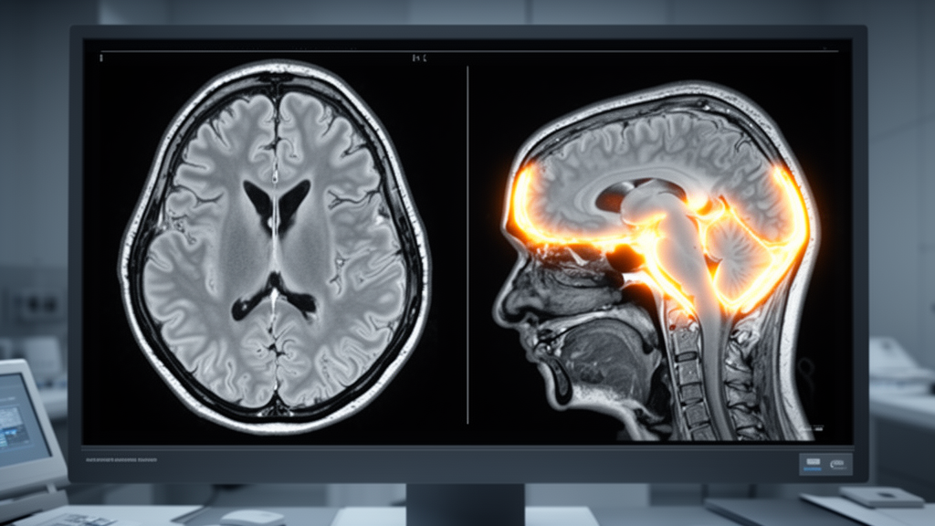
A newly evaluated machine learning framework named SYNEXA combines self-supervised dual-view transformer architectures with post-hoc symbolic explanation mechanisms for multimodal magnetic resonance imaging glioma grading, according to research published in the International Journal of Computational Intelligence Systems. Developed to classify high-grade gliomas and low-grade gliomas, the system prioritizes the empirical validation of internal model evidence alongside diagnostic accuracy. Rather than asserting clinical dominance over established neural network designs, the investigation rigorously quantifies both predictive performance and the interpretive limitations of learned attention maps and surrogate symbolic rules, highlighting critical constraints in automated brain tumor grading workflows.
The architecture processes multimodal brain imaging by constructing paired axial and coronal slabs from four core magnetic resonance imaging sequences: T1-weighted, contrast-enhanced T1-weighted, T2-weighted, and fluid-attenuated inversion recovery scans. To preserve practical clinical applicability, slice selection operates through a mask-free heuristic that identifies slab centers based strictly on fluid-attenuated inversion recovery and contrast-enhanced T1 intensity profiles. Volumetric tumor segmentation masks are deliberately excluded from the feature selection and training pipeline, being reserved exclusively as an evaluation benchmark after training to quantitatively assess model localization behavior against true anatomical tumor boundaries.
Model training is organized across distinct experimental folds using a multi-stage learning schedule. Within each training fold, the dual-view encoder undergoes self-supervised pretraining utilizing masked-patch prediction alongside dual-view contrastive alignment to synchronize complementary spatial representations across the axial and coronal orientations. Following self-supervised initialization, the architecture is fine-tuned using a class-weighted focal loss function optimized on official glioma grade labels from the Multimodal Brain Tumor Segmentation 2020 challenge cohort, balancing learning across asymmetrical diagnostic categories without relying on external annotated guidance.
Across the initial fifteen seed-fold evaluations, SYNEXA demonstrated descriptive mean performance metrics comprising 86.1 percent overall accuracy, 78.6 percent balanced accuracy, a 91.3 percent F1-score, and an area under the receiver operating characteristic curve of 87.4 percent. Because repeated cross-validation creates structural statistical dependencies, the researchers averaged out-of-fold probability outputs across the three iterations for each subject before conducting final patient-level inference. This patient-level validation yielded an estimated accuracy of 85.1 percent with a 95 percent bootstrap confidence interval spanning 81.3 to 88.6 percent, a balanced accuracy of 75.5 percent with a confidence interval between 69.7 and 81.2 percent, an F1-score of 90.7 percent ranging from 88.1 to 93.1 percent, and an area under the curve of 90.4 percent bounded by 86.9 and 93.7 percent.
To evaluate predictive competitiveness under equivalent computational constraints, the authors conducted a controlled benchmark against standard supervised deep learning baselines, training ResNet-18, DenseNet-121, and Vision Transformer ViT-B/16 backbones across an identical or non-smaller maximum budget of 150 supervised epochs per fold. These comparative models achieved descriptive mean accuracies of 86.8 percent, 86.8 percent, and 86.4 percent, respectively. Following paired patient-level testing and Holm multi-comparison correction, no statistically significant difference remained between SYNEXA and any standard baseline. Additionally, the researchers omitted the earlier BraTS 2018 dataset as an external testing benchmark after determining that all 285 of its subjects directly overlapped with the BraTS 2020 cohort.
Spatial interpretability was analyzed through native transformer attention mechanisms, which exhibited modest absolute spatial localization against ground-truth segmentations, recording a Dice-top20 score of 0.218 and a pointing-game score of 0.242. Despite their modest scale, all measured attention metrics significantly exceeded a circular-shift spatial null hypothesis in patient-level statistical testing with a Holm-adjusted p-value of 0.0009. Concurrently, a shallow decision tree surrogate trained on class token embeddings achieved a 90.7 percent fidelity to the primary transformer model. However, the extracted symbolic rules demonstrated extreme cross-run instability, yielding an average Jaccard feature overlap of only 0.005 and indicating that extracted logic reflects model-specific decision paths rather than generalizable clinical criteria.
The study also revealed substantial performance dependencies linked to physical anatomical coverage. The automated slab selection heuristic attained a per-view tumor coverage rate of 64.7 percent. Patient-level diagnostic accuracy reached 92.2 percent when both axial and coronal slabs successfully intersected the tumor lesion, but dropped steeply to 77.8 percent when only a single view contained tumor tissue and fell to 76.5 percent when neither slab captured the lesion. Consequently, the authors position SYNEXA as an auditable, self-supervised classification framework that offers quantitatively audited post-hoc explanations while explicitly cautioning that it should not be viewed as a clinically validated or statistically superior neuro-symbolic medical system.
Subject of Research: Self-supervised dual-view transformer modeling and post-hoc explanation evaluation for multimodal MRI glioma classification
Article Title: SYNEXA: A Self-Supervised Dual-View Transformer with Post-hoc Symbolic Rule Extraction for Multimodal MRI Glioma Classification
Article References: Lakshmi, K. N. M., Nandan, D., & Kumar, S. (2026). SYNEXA: A Self-Supervised Dual-View Transformer with Post-hoc Symbolic Rule Extraction for Multimodal MRI Glioma Classification. International Journal of Computational Intelligence Systems. https://doi.org/10.1007/s44196-026-01683-4
Image Credits: AI Generated
DOI: 10.1007/s44196-026-01683-4
Keywords: SYNEXA, glioma classification, multimodal MRI, dual-view transformer, self-supervised learning, BraTS 2020, HGG and LGG grading, symbolic rule extraction, decision tree surrogate, attention localization, mask-free slice selection, contrastive alignment, masked-patch prediction, cross-validation, model auditability, ResNet-18, DenseNet-121, ViT-B/16
News Source: Cassandra Pierce. (November 18, 2026). Dual-View Brain MRI Transformer Provides Auditable Explanations Without Diagnostic Superiority. Scienmag.