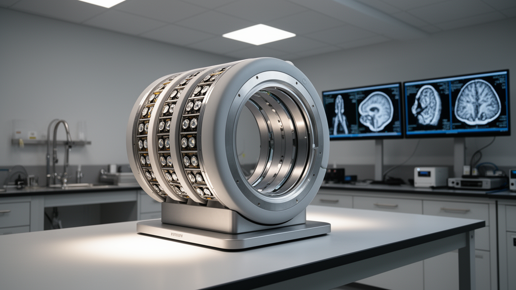
A research team has demonstrated that combining time-of-flight and depth-of-interaction measurements improves brain imaging quality in a dedicated nonhuman primate positron emission tomography scanner.
However, nonhuman primate imaging historically derived limited advantages from time-of-flight information because the smaller physical dimensions of primate subjects offer less spatial localization gain.
To address these simultaneous constraints, the investigators constructed a specialized scanner utilizing an original crosshair light-sharing detector architecture. The detector setup incorporates a 14 by 14 array of fast-LGSO scintillation crystals featuring a 1.6-millimeter pitch and a length of 15 millimeters. The completed scanner ring operates with a compact diameter of 170 millimeters and an axial field of view extending across 156 millimeters.
Instrument performance benchmarks were systematically conducted according to standardized NEMA NU4 and NU2 testing protocols. At a 5-millimeter radial offset from the center, the scanner achieved spatial resolutions of 1.5 millimeters radially, 1.3 millimeters tangentially, and 1.2 millimeters axially.
At an offset distance of 50 millimeters from the scanner center, the radial spatial resolution improved from 4.1 millimeters without interaction depth data down to 2.1 millimeters when depth-of-interaction positioning was engaged.
In evaluations using a sodium-22 multi-rod phantom, individual rods measuring 0.75 millimeters were partially distinguished following iterative image reconstruction. The system demonstrated a peak absolute detection sensitivity of 1.5 percent within an energy acquisition window configured from 440 to 620 kiloelectronvolts.
Count-rate performance assessments employing a monkey-sized scatter phantom showed that the scanner attained a peak noise-equivalent count rate of 65.1 kilocounts per second at an activity level of 102.8 megabecquerels. Furthermore, an in vivo fluorine-18 fluorodeoxyglucose brain scan conducted on a macaque monkey confirmed that incorporating both time-of-flight timing data and depth-of-interaction localization quantitatively improved the visibility of cortical gray matter structures.
The study clarifies that the synergy between depth-of-interaction decoding and time-of-flight registration provides tangible imaging enhancements even within the restricted dimensions of nonhuman primate neuroimaging.
Subject of Research: High-resolution PET imaging system for nonhuman primate neuroimaging
Article Title: A high-resolution TOF-DOI PET system for nonhuman primate neuroimaging
Article References: Ishikawa, T., Akamatsu, G., Ito, S., Mizuma, H., Seki, C., Nagai, Y., Okauchi, T., Wakizaka, H., Iwao, Y., Tashima, H., Yoshida, E., Minamimoto, T., & Yamaya, T. (2026). A high-resolution TOF-DOI PET system for nonhuman primate neuroimaging. EJNMMI Physics. https://doi.org/10.1186/s40658-026-00960-7
Image Credits: AI Generated
DOI: 10.1186/s40658-026-00960-7
Keywords: positron emission tomography, nonhuman primate imaging, time of flight, depth of interaction, neuroimaging, brain PET, fast-LGSO crystals, crosshair light sharing, spatial resolution, macaque monkey, molecular imaging, preclinical neuroscience, PET instrumentation, NEMA standards, scanner design, attenuation and scatter, gray matter visibility
News Source: Cassandra Pierce. (November 13, 2026). Dual TOF and DOI PET System Improves Nonhuman Primate Brain Imaging. Scienmag.