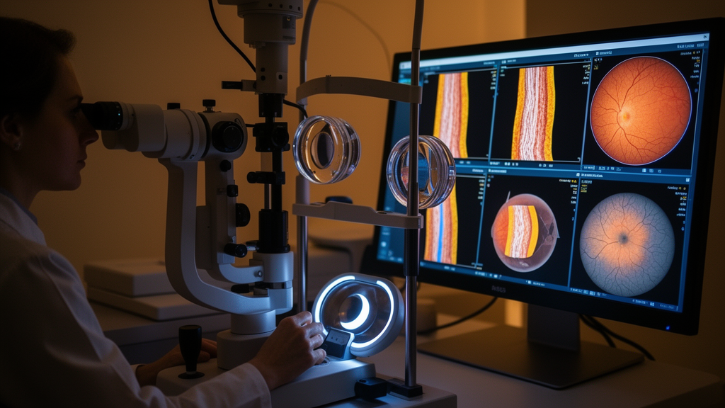
A prospective cohort study has demonstrated that an artificial intelligence framework combining color fundus photography and optical coherence tomography can classify myopia-related fundus pathologies with modality-specific performance. To integrate these complementary perspectives, researchers developed MVFANet, a clinically guided classification framework built around separate pathways tailored to each imaging method.
The study derived its internal dataset from a prospective single-center cohort involving 179 participants. Across this cohort, clinicians collected 4,127 color fundus photography source images from 343 eyes, averaging approximately 12.03 images per eye. In parallel, the investigation gathered 4,526 optical coherence tomography images across 314 eyes, representing an average of 14.41 scans per examined eye. This extensive collection formed the foundational dataset utilized to train and internally validate the independent analytical tracks of the framework.
To assist in training the optical coherence tomography analysis pipeline without imposing rigid runtime data requirements, the researchers incorporated nine-subfield retinal thickness measurements derived directly from corresponding volumetric scans. These structural thickness metrics functioned strictly as auxiliary supervision during the training phase rather than acting as independent inference-time feature inputs.
Model validation followed a rigorous five-fold stratified cross-validation protocol organized strictly at the patient level to eliminate data leakage. All examinations, individual images, and both eyes originating from any single participant were systematically constrained within the same validation fold. Consequently, each participant contributed diagnostic predictions exclusively from the fold in which their clinical data were completely withheld from model parameter updates. Within each training fold, the investigators implemented adaptive loss reweighting to dynamically modulate class-specific error penalties, countering majority-class dominance without exposing out-of-fold data during hyperparameter adjustment.
Internal eye-level diagnostic performance was derived by averaging predicted class probabilities across all images captured from a given eye. Under this evaluation scheme, color fundus photography predictions were finalized by assigning the class with the highest mean probability. In contrast, optical coherence tomography assessments evaluated retinal atrophy and myopic traction independently using fixed decision thresholds. Uncertainty around internal diagnostic metrics was systematically quantified using patient-level clustered bootstrap resampling to generate robust 95% confidence intervals.
Across concatenated out-of-fold predictions, the color fundus photography pathway achieved an internal eye-level classification accuracy of 88.92 percent, with a 95 percent confidence interval ranging from 85.17 to 92.42 percent. This pathway also yielded a Matthews correlation coefficient of 0.8536, a Macro F1 score of 0.8908, and a Cohen’s kappa statistic of 0.8535. For optical coherence tomography evaluations, the framework detected retinal atrophy with an accuracy of 81.85 percent, alongside a Matthews correlation coefficient of 0.6447, a Macro F1 score of 0.8127, and a Cohen’s kappa of 0.6297.
The optical coherence tomography pathway demonstrated higher internal diagnostic metrics when targeting traction lesions, reaching an accuracy of 88.85 percent with a 95 percent confidence interval of 84.59 to 92.85 percent. For this specific pathological feature, the internal validation demonstrated a Matthews correlation coefficient of 0.7518, a Macro F1 score of 0.8726, and a Cohen’s kappa of 0.7461.
External validation on independent public datasets confirmed the generalizability of the dual-modality design following rigorous eligibility screening and standardized regrading based on predefined study criteria. In these external cohorts, the color fundus photography model maintained an accuracy of 84.25 percent and a Macro F1 score of 0.8415. Meanwhile, external optical coherence tomography testing yielded accuracies of 77.71 percent for atrophy and 83.36 percent for traction, corresponding to Macro F1 scores of 0.7703 and 0.8133, respectively.
Grad-CAM-guided occlusion analysis showed larger probability decreases after masking highly activated regions compared with random or low-activation regions, supporting their relevance to model decisions. Despite these findings, the study authors emphasized that prospective integration into clinical decision-support systems requires extensive further validation across broader clinical settings.
Subject of Research: Artificial intelligence assessment of myopia-related fundus pathology using multimodal retinal imaging
Article Title: MVFANet for clinically guided imaging assessment of myopia-related fundus pathology
Article References: Peng, B., Tao, P., Mu, J., Yang, N., Guo, W., Sun, C., Xiang, J., Deng, L., Kang, Y., & Fan, W. (2026). MVFANet for clinically guided imaging assessment of myopia-related fundus pathology. BMC Ophthalmology. https://doi.org/10.1186/s12886-026-05528-8
Image Credits: AI Generated
DOI: 10.1186/s12886-026-05528-8
Keywords: MVFANet, myopia, fundus pathology, color fundus photography, optical coherence tomography, artificial intelligence, deep learning, retinal imaging, retinal atrophy, myopic traction, retinal thickness, auxiliary supervision, stratified cross-validation, external evaluation, Grad-CAM
News Source: Blake Davidson. (November 13, 2026). Dual-Pathway AI Framework Evaluates Myopia Retinal Pathology from Fundus and OCT Imaging. Scienmag.