Researchers at the University of Fukui have demonstrated that electron density imaging derived from dual-energy computed tomography (DECT) can visualize high-grade gliomas in a manner similar to gadolinium-enhanced magnetic resonance imaging (MRI), all without the use of contrast media. Published in Discover Imaging in May 2025, this retrospective study provides preliminary evidence that DECT-based electron density (ED) maps may serve as a viable alternative for patients who cannot undergo MRI due to contraindications such as metal implants, pacemakers, or claustrophobia. The study, led by Takahiro Yamauchi and colleagues, focused on the ability of non-contrast DECT to identify tumor-infiltrating areas and evaluate malignancy in high-grade glioma (HGG) patients.
The central finding of the research is that high electron density areas on DECT images correspond closely to the enhancing areas seen on gadolinium-enhanced T1-weighted MRI (GdT1). In a cohort of ten patients diagnosed with high-grade glioma, the researchers observed a strong positive correlation between the area of high ED on DECT and the enhanced area on GdT1. This suggests that the electron density signature, which reflects the probability of an electron being present at a specific point in space, can highlight regions of high cell density characteristic of aggressive tumors, mimicking the contrast-enhanced appearance traditionally used for diagnosis.
The study was conducted as a single-institution retrospective analysis involving ten patients who underwent both DECT and MRI between January 2021 and October 2023. The cohort consisted of nine patients with glioblastoma and one patient with astrocytoma, grade IV, as defined by the 2021 World Health Organization guidelines. The participants included four males and six females, with a median age of 69.5 years, ranging from 30 to 82 years old. Patients were selected specifically because they presented with noticeable enhanced masses on GdT1 MRI; those with only ring-enhanced masses or no enhancement were excluded to avoid the measurement heterogeneity caused by necrosis and edema.
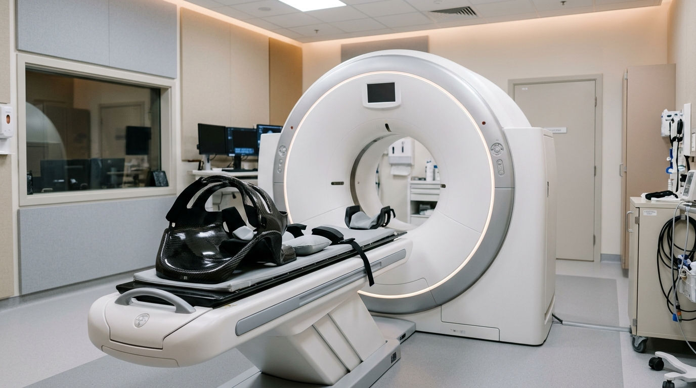
For the imaging protocol, all CT examinations were performed using a dual-source DECT scanner, specifically the SOMATOM Force by Siemens Healthineers. The acquisition utilized a dual-energy mode with tube voltages of 80 kV and 150 kV with tin filtration. The mean CT volume dose index was approximately 65.82 mGy. The MRI scans were obtained using a 3.0-T clinical MRI unit, with standard protocols including plain and gadolinium-enhanced T1-weighted sequences. The time interval between the DECT and MRI evaluations was limited to two weeks to ensure comparability of the tumor status.
Data processing involved smoothing the ED images using 3D median and Gaussian blur filters to reduce noise, followed by the application of a color lookup table to emphasize the tumor regions. Three raters, including two board-certified neurosurgeons and one radiologist, independently analyzed the images. They measured the area of high ED on the DECT images and the enhanced area on the GdT1 images, correcting for differences in brain size between the two modalities by measuring the length and width of the brain on the same slice. The high ED area was defined visually as having a density higher than that of the contralateral structure.
Statistical analysis revealed a significant positive correlation between the area ratios of high ED and Gd enhancement across all raters. Linear regression analysis showed R-squared values of 0.910, 0.857, and 0.717 for the three raters, with p-values less than 0.0001, 0.0001, and 0.002, respectively. The intraclass correlation coefficient (ICC) for the ratio of area ratios was 0.75, indicating good inter-rater reliability. These results suggest that the visualization of high ED areas is consistent among experienced medical professionals, despite the subjective nature of the initial visual definition.
Furthermore, the study compared the relative electron density (rED) of the tumor area with that of the contralateral white matter (CLWM). The rED was calculated using a formula based on the Hounsfield unit difference relative to water. The mean and maximum rED of the tumor area were significantly higher than those of the CLWM, with p-values of 0.049 and 0.0002, respectively. In contrast, the minimum rED did not show a significant difference between the tumor and CLWM. This indicates that the peak electron density within the tumor is a more reliable marker for distinguishing high-grade glioma tissue from normal brain parenchyma.
Receiver operating characteristic (ROC) curve analysis provided additional diagnostic metrics. For the maximum rED, the area under the curve (AUC) was 0.962, with a cut-off value of 1.031. This metric achieved a sensitivity of 100% and a specificity of 77.8%, along with a positive predictive value of 81.8% and a negative predictive value of 100%. The mean rED also showed diagnostic utility with an AUC of 0.802, while the minimum rED had a lower AUC of 0.654. These figures suggest that the maximum rED is the most effective single metric for differentiating high-grade glioma from normal white matter in this specific cohort.
Pathological correlation in representative cases supported the imaging findings. In a biopsy case, specimens from the high ED and Gd-enhanced areas showed high cell density and a Ki-67 proliferation index of 10%. In a lobectomy case, the high ED area corresponded to tissue with high cell density and a Ki-67 index of 32%, whereas the low ED area showed low cell density and a Ki-67 index of 5%. This histological alignment reinforces the hypothesis that electron density reflects cellular density, which is a key feature of malignancy in gliomas.
The authors acknowledge several limitations of the study. The sample size was small, consisting of only ten patients, which limits the generalizability of the findings. The study included only patients with noticeable enhanced masses, excluding those with ring-enhancing patterns or cysts, which are common in high-grade gliomas. Additionally, the relationship between rED and cell density has not been verified against established methods like methionine-PET. The authors note that further research is needed to clarify the mechanism of higher ED near bone and to include a wider variety of tumor shapes in future studies.
Despite these limitations, the study offers a promising avenue for patients who cannot undergo MRI. MRI is the gold standard for glioma diagnosis but is inaccessible to patients with certain implants or conditions. DECT is generally less expensive and faster than MRI, and the use of electron density imaging eliminates the need for gadolinium contrast, which carries risks of allergy and renal dysfunction. The ability to visualize high-density tumor areas without contrast could expand diagnostic options and improve preoperative planning for a subset of high-grade glioma patients.
This research contributes to the growing body of evidence supporting the use of advanced CT techniques in neuro-oncology. By demonstrating that non-contrast DECT can mimic the diagnostic utility of contrast-enhanced MRI for high-grade gliomas, the study highlights the potential of electron density as a biomarker for tumor malignancy. Future studies with larger cohorts and diverse tumor morphologies will be essential to validate these findings and determine the clinical applicability of this technique in routine practice.
Subject of Research: Comparison of electron density imaging from dual-energy CT and MRI for high-grade glioma diagnosis
Article Title: Comparison between electron density imaging with dual-energy computed tomography without contrast medium and magnetic resonance imaging for high-grade glioma
Article References: Yamauchi, T., Ishida, T., Itoh, T., Tsubota, T., Takata, K., Higashino, Y., Tsujikawa, T., & Kikuta, K.-I. (2025). Comparison between electron density imaging with dual-energy computed tomography without contrast medium and magnetic resonance imaging for high-grade glioma. Discover Imaging, 2(1), Article 4. https://doi.org/10.1007/s44352-025-00008-z
Image Credits: AI Generated
DOI: 10.1007/s44352-025-00008-z
Keywords: dual-energy CT, high-grade glioma, electron density, MRI, neuro-oncology, contrast-free imaging, glioblastoma, medical imaging, tumor diagnosis, Comparison, between, electron
News Source: Ophelia Keating. (November 11, 2026). Dual-Energy CT Visualizes High-Grade Glioma Without Contrast. Scienmag.