


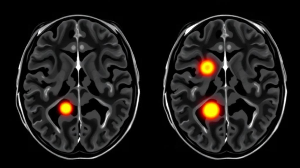
Double inversion recovery (DIR) magnetic resonance imaging detected more cervical spinal cord lesions in people with multiple sclerosis than the two sequences routinely used in clinical practice, according to a cross-sectional study of 100 patients scanned at 3 Tesla. In a cohort pre-selected for confirmed spinal cord involvement, DIR identified lesions in every patient, while short tau inversion recovery (STIR) missed one patient and conventional T2-weighted imaging missed four. The findings, published in the Egyptian Journal of Radiology and Nuclear Medicine, suggest that DIR could earn a place alongside existing sequences when imaging the cervical cord, a region where small anatomy, motion and cerebrospinal fluid make lesion detection notoriously difficult.
The numbers behind that headline are striking. Across the 100 patients, DIR yielded a mean of 5.02 lesions per person, with a standard deviation of 2.89, for a total of 389 lesions. STIR detected a mean of 4.58 lesions per patient, totaling 361, and T2-weighted imaging detected a mean of 3.67, totaling 290. The differences were statistically significant, with a p value below 0.001 and a partial eta-squared of 0.38, which the authors describe as a large effect size. Detection rates within this pre-selected cohort were 100 percent for DIR (95 percent confidence interval 96.4 to 100 percent), 99 percent for STIR and 96 percent for T2-weighted imaging. Because every participant already had confirmed cervical cord lesions, these rates describe how well each sequence visualized disease that was known to exist, not how well the sequences screen an unselected population.
Conspicuity told a similar story. DIR showed a striking predominance of marked intensity, at 65 percent of lesions. STIR lesions were predominantly moderate, at 58 percent, while T2-weighted imaging showed predominantly mild intensity, at 81 percent. The distribution of intensities differed significantly across the three sequences. In representative images, lesions that appeared as two small, clearly demarcated hyperintense plaques on DIR showed up as a single faint plaque on STIR and were entirely invisible on T2-weighted imaging at the same vertebral level.
DIR achieves this contrast through a fundamentally different mechanism. Standard fluid-attenuated inversion recovery applies a single inversion pulse to suppress only cerebrospinal fluid. DIR applies two distinct inversion pulses, simultaneously suppressing signal from both cerebrospinal fluid and normal white matter. That dual suppression renders gray matter and inflammatory lesions hyperintense against a darkened background, sharpening the contrast between pathology and healthy tissue. The technique is particularly valuable where partial volume effects with cerebrospinal fluid can obscure pathology, a persistent problem in the narrow cervical cord. In this study the sequence used a short inversion time of 270 to 340 milliseconds and a long inversion time of 3000 to 3400 milliseconds, with a parallel imaging acceleration factor of 2 and a relatively short total acquisition time of 3 minutes and 45 seconds.
The study enrolled patients attending neurology outpatient clinics at a single institution between May 2023 and April 2024, following STROBE reporting guidelines for observational studies. Participants were aged up to 60 years and fulfilled the 2017 revised McDonald criteria together with the 2021 MAGNIMS guidelines. The cohort was predominantly female, at 63 percent, with a mean age of 33.71 years. Relapsing-remitting disease accounted for 85 percent of patients, secondary progressive disease for 14 percent and primary progressive disease for 1 percent. Mean disease duration was 6.00 years, mean age at onset was 27.14 years, and the mean Expanded Disability Status Scale score was 2.96. Motor involvement was reported in 75 percent of patients and sensory involvement in 76 percent, with visual, brainstem, cerebellar and sphincteric involvement also common.
The imaging comparison rested on careful reading protocols. Two experienced neuroradiologists, with 10 and 8 years of MS MRI experience respectively, first reviewed clinical records and available examinations to confirm eligibility and exclude confounding abnormalities such as spinal cord trauma or advanced degenerative disk disease. They then independently interpreted the three compared sequences, blinded to one another’s readings, assessing lesion existence, number, anatomical level and conspicuity. A lesion was considered confirmed when it appeared as a focal hyperintense abnormality on at least two of the three sequences, and disagreements were resolved in a consensus session that served as the reference standard. Interobserver agreement was excellent, with a Cohen’s kappa of 0.89 for lesion detection and intensity grading and an intraclass correlation coefficient of 0.92 for lesion counts, with a 95 percent confidence interval of 0.88 to 0.95.
The authors place their results in the context of a literature that has long celebrated DIR in the brain but rarely tested it in the spine. Introduced by Redpath and Smith in 1994, the sequence has shown exceptional sensitivity for MS lesions in the brain, especially intracortical lesions, and one prior study reported that DIR outperformed T2-weighted imaging across periventricular, mixed white-gray matter, cortical and infratentorial regions, with relative gains of 31, 47, 98 and 39 percent and an overall 18 percent gain in total lesion load. For the cervical spine specifically, earlier work reported DIR sensitivity of 87 percent for lesions, a 37 percent increase in lesions detected compared with FLAIR, and higher lesion-to-noise ratios. Another group found that reformatted DIR images detected 115 lesions versus 96 on conventional T2-weighted turbo spin echo images, an overall 28 percent increase, averaging 4 lesions per patient against 3. Not all studies agree, however; one reported that volumetric DIR detected the fewest lesions, a discrepancy the authors attribute to acquisition parameters, noting that their own sequence used the shortest echo train time, 97 milliseconds, and echo time, 200 milliseconds, among the compared DIR studies.
Beyond detection, the study explored whether DIR lesion burden relates to clinical status, and the answer is a qualified yes. The number of DIR-detected lesions correlated positively with disease progression, defined as a confirmed increase of at least 1.0 EDSS point sustained over at least 3 months, with a Spearman coefficient of 0.369 and a p value below 0.001. DIR lesion count also correlated with EDSS score, with a coefficient of 0.215 and a p value of 0.041. The authors are careful to stress that these are weak correlations, explaining only 13.6 percent and 4.6 percent of the variance respectively, and that DIR cannot be considered a standalone predictor of clinical disability. This modest association echoes the long-recognized clinico-radiological paradox in multiple sclerosis, in which MRI lesion load correlates poorly with disability, and prior work has shown that some small DIR-visible cortical lesions do not track with clinical symptoms.
Notably, no significant correlations emerged between MRI findings and disease activity, nor with demographic variables including age, sex, age of onset, disease type, disease duration or relapse frequency. The authors attribute the disease activity result in part to the absence of gadolinium-enhanced T1-weighted imaging, the established standard for identifying active inflammatory lesions, meaning no conclusions about DIR’s utility for monitoring disease activity can be drawn from this study. They also acknowledge that DIR is more technically demanding than its rivals: the sequence’s long repetition time and dual inversion pulses make it susceptible to motion artifacts from cerebrospinal fluid pulsation and swallowing. The team mitigated this with the short acquisition time, careful patient positioning and a technologist present throughout scanning, and excluded patients with significant motion artifact or poor image quality.
The limitations frame how far these conclusions can travel. The single-center design, the relatively homogeneous sample dominated by relapsing-remitting disease, and the consensus-based imaging reference standard, which involved no histopathological verification, all temper the findings. The reported detection rates therefore describe performance relative to expert consensus rather than definitive ground truth, and the authors recommend future multicenter studies with larger and more diverse populations, ideally incorporating full-coverage axial T2-weighted imaging to confirm lesions and assess their cross-sectional characteristics. Within those bounds, the message is concrete: at 3 Tesla, with optimized parameters, double inversion recovery visualized more cervical cord lesions, and visualized them more conspicuously, than either STIR or conventional T2-weighted imaging in the same patients, supporting its evaluation as an adjunctive sequence in cervical cord MRI protocols for multiple sclerosis.
Subject of Research: Double inversion recovery MRI for detecting cervical spinal cord lesions in multiple sclerosis
Article Title: 3-Tesla double inversion recovery MRI for improved detection of cervical cord lesions in multiple sclerosis: a cross-sectional study
Article References: Difallah, D. K., Ghalab, M., Ragab, S., & Foda, H. M. (2026). 3-Tesla double inversion recovery MRI for improved detection of cervical cord lesions in multiple sclerosis: a cross-sectional study. Egyptian Journal of Radiology and Nuclear Medicine, 57(1), Article 217. https://doi.org/10.1186/s43055-026-01865-2
Image Credits: AI Generated
DOI: 10.1186/s43055-026-01865-2
Keywords: multiple sclerosis, double inversion recovery, MRI, cervical spinal cord, STIR, T2-weighted imaging, lesion detection, 3 Tesla, neuroradiology, EDSS, cross-sectional study, demyelination
News Source: Cassandra Pierce. (October 29, 2026). Double Inversion Recovery MRI Spots More Spinal Cord Lesions in Multiple Sclerosis. Scienmag.



