


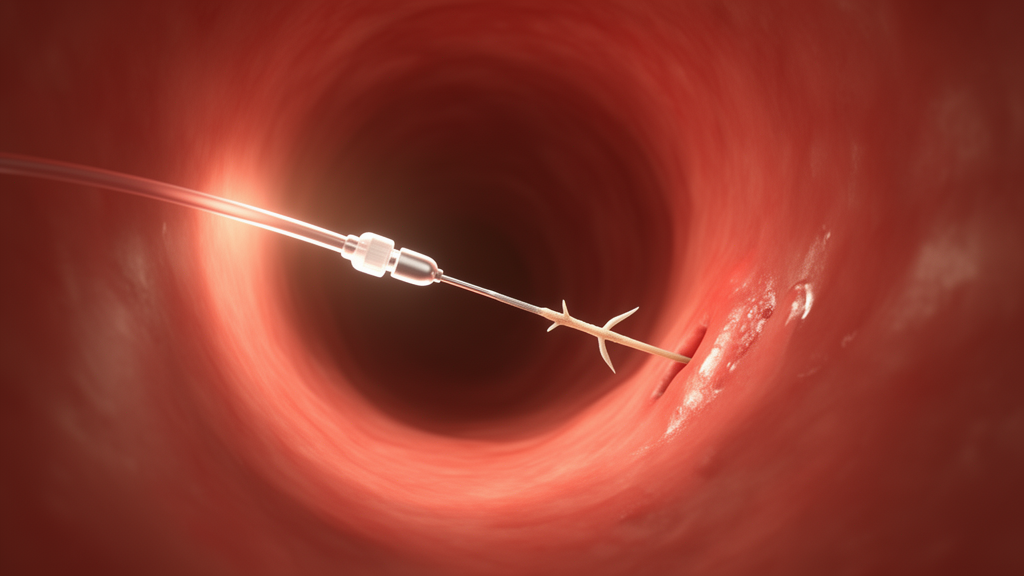
A swallowed fish bone that migrated into one of the largest veins in the human body has been successfully removed using intravascular ultrasound guidance, according to a case report published in CVIR Endovascular. The patient, a 70-year-old woman, arrived at an outpatient clinic with a one-month history of night sweats, chills, and pain in her right iliac fossa. She was undergoing gemcitabine chemotherapy for stage IV cholangiocarcinoma, and earlier courses of antibiotics prescribed for a suspected urinary tract infection and gingival inflammation had failed to relieve her symptoms.
Contrast-enhanced CT performed for oncologic follow-up revealed focal nonocclusive thrombosis of the infrarenal inferior vena cava containing a linear hyperdense foreign body compatible with a fish bone. One extremity of the object extended into a small right renal vein. The scan also showed marked inflammatory fat stranding between the aorta and the cava, adjacent to the third portion of the duodenum, which the authors interpreted as the presumed route by which the bone had eroded through the bowel wall and entered the vein. A small segmental pulmonary embolism was present as well, visible in the right lower lobe on CT pulmonary angiography. Only after these findings did the patient recall having recently increased her fish consumption, though she could not remember swallowing a bone or experiencing any choking episode.
Intravascular migration of ingested fish bones is exceptionally rare. Most ingested foreign bodies pass spontaneously through the gastrointestinal tract without clinical consequences, and gastrointestinal perforation occurs in fewer than 1% of cases. The estimated incidence of fish bone–related bowel perforation is approximately 2.9 per 100,000 person-years. When perforation does occur, sharp objects such as fish bones may migrate into adjacent structures and cause severe vascular complications including thrombosis, pseudoaneurysm formation, hemorrhage, and septic embolization. A review of 107 cases of migrating fish bones and toothpicks found that the peritoneal cavity was the most common destination, accounting for 64.5% of cases, which underscores how unusual vascular involvement is. Venous involvement in particular is distinctly uncommon and often presents with nonspecific symptoms, making diagnosis challenging.
The patient was admitted and started on intravenous piperacillin–tazobactam, daptomycin, and therapeutic anticoagulation. Despite broad-spectrum intravenous antibiotics, she continued to experience malaise, night sweats, and chills, with persistent leukocytosis and elevated C-reactive protein levels. Given the persistent systemic inflammatory syndrome, the presence of pulmonary embolism, and the retained intravascular foreign body with its ongoing risk of infectious and thromboembolic complications, her team concluded that conservative management alone was insufficient. After a multidisciplinary discussion, they proceeded to endovascular retrieval, a minimally invasive alternative to open surgery.
The procedure revealed exactly why this case was technically demanding. Initial venography of the inferior vena cava with digital subtraction angiography, performed through right femoral venous access, demonstrated only a subtle filling defect. Even with digital subtraction, the foreign body remained poorly visualized under fluoroscopy, becoming barely perceptible only after contrast injection. Intravascular ultrasound told a completely different story: it clearly showed a hyperechogenic linear structure with posterior acoustic shadowing embedded within focal caval thrombosis and extending toward a renal vein. The ultrasound catheter allowed the team to identify the free intraluminal tip of the bone, the critical detail that made targeted retrieval possible.
To reduce the risk of embolization during manipulation, the team deployed a temporary suprarenal inferior vena cava filter through an 8-Fr right internal jugular vein access. Under systemic anticoagulation with intravenous heparin, with an activated clotting time of 200 seconds, an OptiCross 35 intravascular ultrasound catheter was advanced through the jugular access to localize the foreign body and assess its orientation relative to the surrounding thrombus. After upsizing the femoral venous access to a 12-Fr sheath, retrieval was first attempted with a 20-mm single-loop snare, then a triple-loop snare, and finally a handmade snare fashioned from a 0.035-inch guidewire advanced through a Simmons diagnostic catheter. None of these techniques successfully engaged the bone, and ultrasound confirmed it had not been displaced by the repeated attempts.
The successful solution came from an off-label tool: a rat-tooth biopsy forceps, a device normally used in endoscopy. Introduced through the sheath, the forceps were operated under combined fluoroscopic and real-time intravascular ultrasound guidance. The free intraluminal tip of the foreign body was grasped and removed, and the retrieved object was confirmed to be a fish bone. Final ultrasound evaluation demonstrated residual mural caval thrombosis without procedural complications, and completion venography showed no residual filling defects. The protective filter was then retrieved. The authors note that although loop snares remain the most commonly used retrieval devices, their effectiveness can be limited by the morphology, orientation, and degree of embedding of a foreign body, and that biopsy forceps have demonstrated high technical success and low complication rates in large series of inferior vena cava filter retrieval.
The main risk of grasping forceps inside a major vein is inadvertent capture of the venous wall, with potential perforation or pseudoaneurysm formation. In this case, the authors write, that risk was mitigated by real-time ultrasound confirmation of the free intraluminal tip before grasping, and by careful manipulation through a 12-Fr sheath under continuous combined ultrasound and fluoroscopic guidance. The day after the intervention, the patient reported complete resolution of her symptoms. She was discharged asymptomatic after completing the prescribed antibiotic course, with no procedure-related complications, and was maintained on therapeutic oral anticoagulation for a planned total duration of 6 months. At one-month follow-up, CT demonstrated complete resolution of the inferior vena cava thrombosis and the surrounding inflammatory changes, and no further intervention related to the foreign body was deemed necessary. She continued routine oncologic follow-up for her underlying malignancy while remaining asymptomatic from the vascular complication.
The authors emphasize that intravascular ultrasound provided a major procedural advantage by allowing real-time visualization of a foreign body that was nearly invisible on fluoroscopy, enabling precise localization, assessment of its relationship to the vessel wall and thrombus, and potentially reducing the risk of embolization or vascular injury. Compared with previously published reports, which predominantly describe arterial complications or fluoroscopy-guided retrieval, this case stands out for its venous involvement of the inferior vena cava with extension into a renal vein, associated thrombosis and pulmonary embolism, and successful retrieval using endoscopic forceps. The team also notes that surgical removal would likely have been the next option had percutaneous techniques failed, since conservative management would have left the bone in place with persistent risks of infection, thromboembolic complications, and ongoing inflammation.
As a single case report, the findings carry important limitations. The authors state that it does not allow comparison with alternative management strategies or establish the superiority of ultrasound-guided retrieval over conventional fluoroscopic techniques. Given the extreme rarity of intravascular fish bone migration, they conclude that multicenter registries and additional case reports will be required to better define patient selection, procedural strategies, and long-term outcomes. For now, the case illustrates a practical lesson for interventional teams: when a foreign body hides within venous thrombus and fluoroscopy fails, ultrasound delivered from inside the vessel can light the way, and an endoscopic biopsy forceps can serve as an effective bailout when conventional snares cannot get a grip.
Subject of Research: Intravascular ultrasound-guided endovascular retrieval of a migrated fish bone causing inferior vena cava thrombosis
Article Title: Intravascular ultrasound-guided retrieval of a fish bone embedded in the inferior vena cava: a case report
Article References: Conde, L. F., Matute-González, M., Gelabert, A., Sueiras, A., & Barrufet, M. (2026). Intravascular ultrasound-guided retrieval of a fish bone embedded in the inferior vena cava: a case report. CVIR Endovascular, 9(1), Article 125. https://doi.org/10.1186/s42155-026-00778-5
Image Credits: AI Generated
DOI: 10.1186/s42155-026-00778-5
Keywords: intravascular ultrasound, fish bone, inferior vena cava, venous thrombosis, pulmonary embolism, endovascular retrieval, foreign body migration, case report, biopsy forceps, interventional radiology, IVC filter, gastrointestinal perforation
News Source: Ophelia Keating. (October 29, 2026). Doctors Use Ultrasound Inside a Vein to Pull a Swallowed Fish Bone Out of the Inferior Vena Cava. Scienmag.



